1PXD image
Entry Detail
PDB ID:
1PXD
Title:
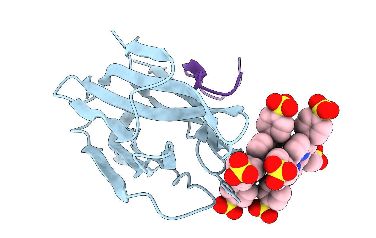
Crystal structure of the complex of jacalin with meso-tetrasulphonatophenylporphyrin.
Biological Source:
Source Organism:
PDB Version:
Deposition Date:
2003-07-03
Release Date:
2004-02-03
Method Details:
Experimental Method:
Resolution:
1.80 Å
R-Value Free:
0.23
R-Value Work:
0.21
R-Value Observed:
0.21
Space Group:
I 2 2 2
Macromolecular Entities
Polymer Type:polypeptide(L)
Description:Agglutinin alpha chain
Chain IDs:A
Chain Length:133
Number of Molecules:1
Biological Source:Artocarpus integer
Polymer Type:polypeptide(L)
Description:Agglutinin beta-3 chain
Chain IDs:B
Chain Length:20
Number of Molecules:1
Biological Source:Artocarpus integer
Ligand Molecules
Primary Citation
Porphyrin binding to jacalin is facilitated by the inherent plasticity of the carbohydrate-binding site: novel mode of lectin-ligand interaction.
Acta Crystallogr.,Sect.D 60 281 288 (2004)
PMID: 14747704 DOI: 10.1107/S0907444903026684

Abstact

The crystal structure of the complex of meso-tetrasulfonatophenylporphyrin (H(2)TPPS) with jack fruit (Artocarpus integriflora) agglutinin (jacalin) has been determined at 1.8 A resolution. A porphyrin pair is sandwiched between two symmetry-related jacalin monomers in the crystal, leading to a cross-linking network of protein molecules. Apart from the stacking interactions, H(2)TPPS also forms hydrogen bonds, some involving water bridges, with jacalin at the carbohydrate-binding site. The residues that are involved in rendering galactopyranoside specificity to jacalin undergo conformational adjustments in order to accommodate the H(2)TPPS molecule. The water molecules at the carbohydrate-binding site of jacalin cement the jacalin-porphyrin interactions, optimizing their complementarity. Interactions of porphyrin with jacalin are relatively weak compared with those observed between galactopyranoside and jacalin, perhaps because the former largely involves water-mediated hydrogen bonds. While H(2)TPPS binds to jacalin at the carbohydrate-binding site as in the case of ConA, its mode of interaction with jacalin is very different. H(2)TPPS does not enter the carbohydrate-binding cavity of jacalin. Instead, it sits over the binding site. While the porphyrin binding is mediated by replicating the hydrogen-bonding network of mannopyranoside through the sulfonate atoms in the case of ConA, the plasticity associated with the carbohydrate-binding site accommodates the pluripotent porphyrin molecule in the case of jacalin through an entirely different set of interactions.

Legend

Protein

Chemical

Disease

Primary Citation of related structures