1PVE image
Deposition Date 2003-06-27
Release Date 2004-08-10
Last Version Date 2024-05-29
Entry Detail
PDB ID:
1PVE
Title:
Solution structure of XPC binding domain of hHR23B
Biological Source:
Source Organism(s):
Homo sapiens (Taxon ID: 9606)
Expression System(s):
Method Details:
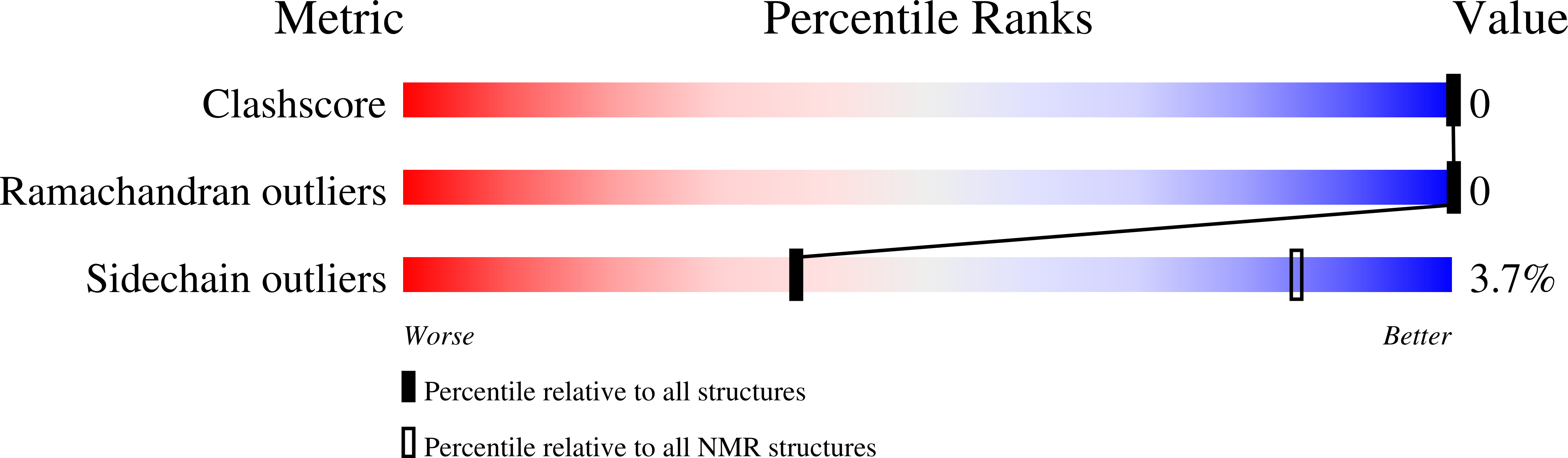
Experimental Method:
Conformers Calculated:
50
Conformers Submitted:
20
Selection Criteria:
structures with the least restraint violations
Macromolecular Entities
Structures with similar UniProt ID
Protein Blast
Polymer Type:polypeptide(L)
Molecule:UV excision repair protein RAD23 homolog B
Gene (Uniprot):RAD23B
Chain IDs:A
Chain Length:72
Number of Molecules:1
Biological Source:Homo sapiens
Ligand Molecules
Primary Citation
Solution structure and backbone dynamics of the XPC-binding domain of the human DNA repair protein hHR23B.
Febs J. 272 2467 2476 (2005)
PMID: 15885096 DOI: 10.1111/j.1742-4658.2005.04667.x

Abstact

Human cells contain two homologs of the yeast RAD23 protein, hHR23A and hHR23B, which participate in the DNA repair process. hHR23B houses a domain (residues 277-332, called XPCB) that binds specifically and directly to the xeroderma pigmentosum group C protein (XPC) to initiate nucleotide excision repair (NER). This domain shares sequence homology with a heat shock chaperonin-binding motif that is also found in the stress-inducible yeast phosphoprotein STI1. We determined the solution structure of a protein fragment containing amino acids 275-342 of hHR23B (termed XPCB-hHR23B) and compared it with the previously reported solution structures of the corresponding domain of hHR23A. The periodic positioning of proline residues in XPCB-hHR23B produced kinked alpha helices and assisted in the formation of a compact domain. Although the overall structure of the XPCB domain was similar in both XPCB-hHR23B and XPCB-hHR23A, the N-terminal part (residues 275-283) of XPCB-hHR23B was more flexible than the corresponding part of hHR23A. We tried to infer the characteristics of this flexibility through (15)N-relaxation studies. The hydrophobic surface of XPCB-hHR23B, which results from the diverse distribution of N-terminal region, might give rise to the functional pleiotropy observed in vivo for hHR23B, but not for hHR23A.

Legend

Protein

Chemical

Disease

Primary Citation of related structures
Feedback Form
Name
Email
Institute
Feedback