Deposition Date
2003-06-26
Release Date
2004-05-25
Last Version Date
2024-05-01
Entry Detail
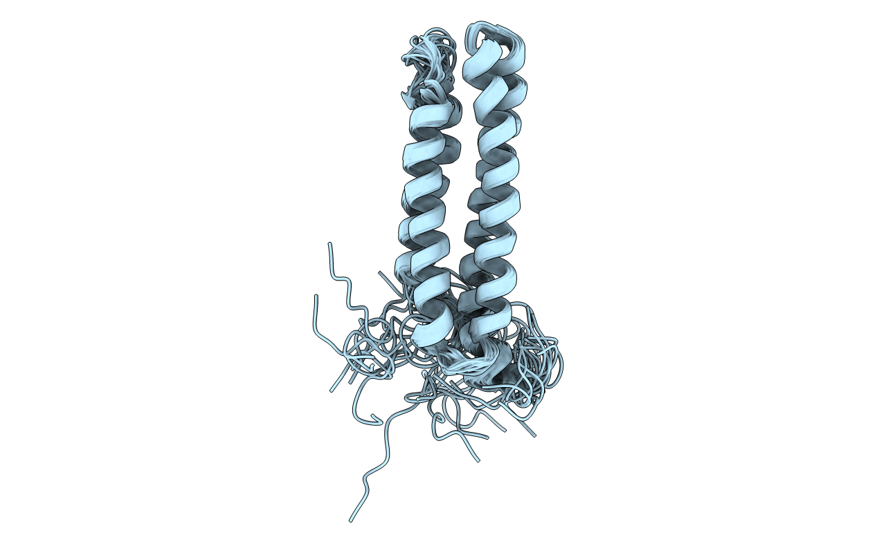
PDB ID:
1PV3
Keywords:
Title:
NMR Solution Structure of the Avian FAT-domain of Focal Adhesion Kinase
Biological Source:
Source Organism(s):
Gallus gallus (Taxon ID: 9031)
Expression System(s):
Method Details:
Experimental Method:
Conformers Calculated:
25
Conformers Submitted:
20
Selection Criteria:
structures with acceptable covalent geometry,structures with the lowest energy