
Deposition Date
1995-04-21
Release Date
1996-08-01
Last Version Date
2024-11-20
Entry Detail
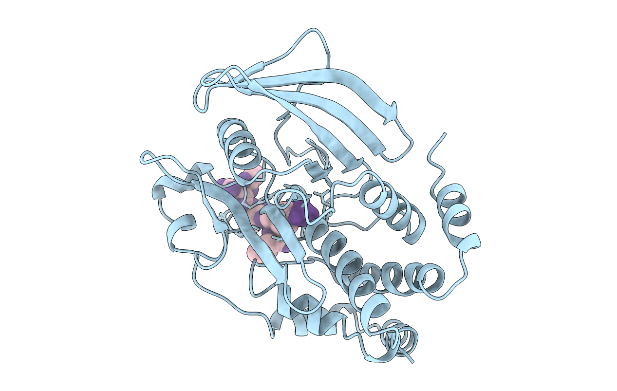
PDB ID:
1PTT
Keywords:
Title:
CRYSTAL STRUCTURE OF PROTEIN TYROSINE PHOSPHATASE 1B COMPLEXED WITH PHOSPHOTYROSINE-CONTAINING TETRA-PEPTIDE (AC-DEPYL-NH2)
Biological Source:
Source Organism(s):
Homo sapiens (Taxon ID: 9606)
Expression System(s):
Method Details:
Experimental Method:
Resolution:
2.90 Å
R-Value Work:
0.19
R-Value Observed:
0.19
Space Group:
P 31 2 1
