1PSF image
Deposition Date 1994-04-13
Release Date 1995-04-20
Last Version Date 2024-05-01
Entry Detail
PDB ID:
1PSF
Keywords:
Title:
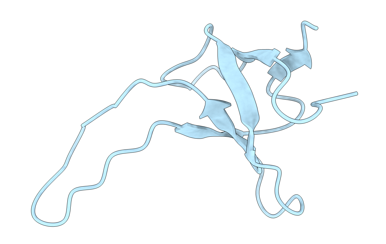
THE THREE-DIMENSIONAL SOLUTION STRUCTURE OF PSAE FROM THE CYANOBACTERIUM SYNECHOCOCCUS SP. STRAIN PCC 7002: A PHOTOSYSTEM I PROTEIN THAT SHOWS STRUCTURAL HOMOLOGY WITH SH3 DOMAINS
Biological Source:
Source Organism:
Method Details:
Experimental Method:
Conformers Submitted:
1
Macromolecular Entities
Polymer Type:polypeptide(L)
Molecule:PHOTOSYSTEM I ACCESSORY PROTEIN E
Gene (Uniprot):psaE
Chain IDs:A
Chain Length:69
Number of Molecules:1
Biological Source:Synechococcus sp.
Ligand Molecules
Primary Citation
Three-dimensional solution structure of PsaE from the cyanobacterium Synechococcus sp. strain PCC 7002, a photosystem I protein that shows structural homology with SH3 domains.
Biochemistry 33 6052 6062 (1994)
PMID: 8193119 DOI: 10.1021/bi00186a004

Abstact

PsaE is a 69 amino acid polypeptide from photosystem I present on the stromal side of the thylakoid membrane. The three-dimensional solution structure of this protein from the cyanobacterium Synechococcus sp. strain PCC 7002 was determined at pH 5.8 and room temperature using over 900 experimental restraints derived from two- and three-dimensional NMR experiments. The structure is comprised of a well-defined five-stranded beta-sheet with (+1, +1, +1, -4 alpha) topology. There is no helical region except for a single turn of 3(10) helix between the beta D and beta E strands. PsaE also exhibits a large unrestrained loop spanning residues 42-56. A comparison to known protein structures revealed similarity with the Src homology 3 (SH3) domain, a membrane-associated protein involved in signal transduction in eukaryotes. The match is remarkable as 47 of the alpha-carbons of PsaE can be superimposed onto those of the SH3 domain from chicken brain alpha-spectrin with a root-mean-square deviation of 2.3 A. Although the amino acid sequences have low identity and the loops are different in both proteins, the topology of the beta-sheet and the 3(10) turn is conserved. SH3 domains from other sources show a similar structural homology. The structure of PsaE was used to suggest approaches for elucidating its roles within photosystem I.

Legend

Protein

Chemical

Disease

Primary Citation of related structures