1PRZ image
Deposition Date 2003-06-20
Release Date 2003-11-04
Last Version Date 2024-11-06
Entry Detail
PDB ID:
1PRZ
Keywords:
Title:
Crystal structure of pseudouridine synthase RluD catalytic module
Biological Source:
Source Organism:
Escherichia coli (Taxon ID: 562)
Host Organism:
Method Details:
Experimental Method:
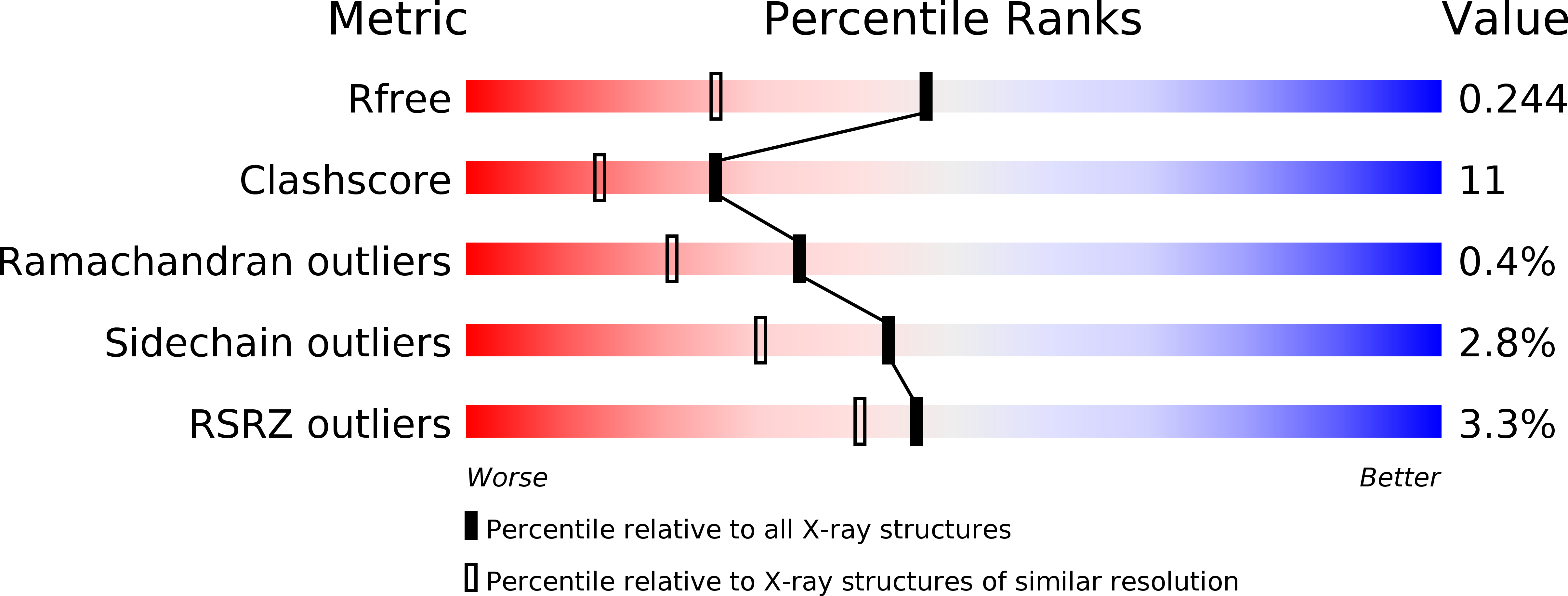
Resolution:
1.80 Å
R-Value Free:
0.24
R-Value Work:
0.21
Space Group:
P 21 21 21
Macromolecular Entities
Polymer Type:polypeptide(L)
Molecule:Ribosomal large subunit pseudouridine synthase D
Chain IDs:A
Chain Length:252
Number of Molecules:1
Biological Source:Escherichia coli
Modified Residue
Compound ID Chain ID Parent Comp ID Details 2D Image
MSE A MET SELENOMETHIONINE
Primary Citation
Crystal structure of the RluD pseudouridine Synthase catalytic module, an enzyme that modifies 23S rRNA and is essential for normal cell growth of Escherichia coli
J.Mol.Biol. 335 87 101 (2003)
PMID: 14659742 DOI: 10.1016/j.jmb.2003.10.003

Abstact

Pseudouridine (5-beta-D-ribofuranosyluracil, Psi) is the most commonly found modified base in RNA. Conversion of uridine to Psi is performed enzymatically in both prokaryotes and eukaryotes by pseudouridine synthases (EC 4.2.1.70). The Escherichia coli Psi-synthase RluD modifies uridine to Psi at positions 1911, 1915 and 1917 within 23S rRNA. RluD also possesses a second function related to proper assembly of the 50S ribosomal subunit that is independent of Psi-synthesis. Here, we report the crystal structure of the catalytic module of RluD (residues 68-326; DeltaRluD) refined at 1.8A to a final R-factor of 21.8% (R(free)=24.3%). DeltaRluD is a monomeric enzyme having an overall mixed alpha/beta fold. The DeltaRluD molecule consists of two subdomains, a catalytic subdomain and C-terminal subdomain with the RNA-binding cleft formed by loops extending from the catalytic sub-domain. The catalytic sub-domain of DeltaRluD has a similar fold as in TruA, TruB and RsuA, with the location of the RNA-binding cleft, active-site and conserved, catalytic Asp residue superposing in all four structures. Superposition of the crystal structure of TruB bound to a T-stem loop with RluD reveals that similar RNA-protein interactions for the flipped-out uridine base would exist in both structures, implying that base-flipping is necessary for catalysis. This observation also implies that the specificity determinants for site-specific RNA-binding and recognition likely reside in parts of RluD beyond the active site.

Legend

Protein

Chemical

Disease

Primary Citation of related structures