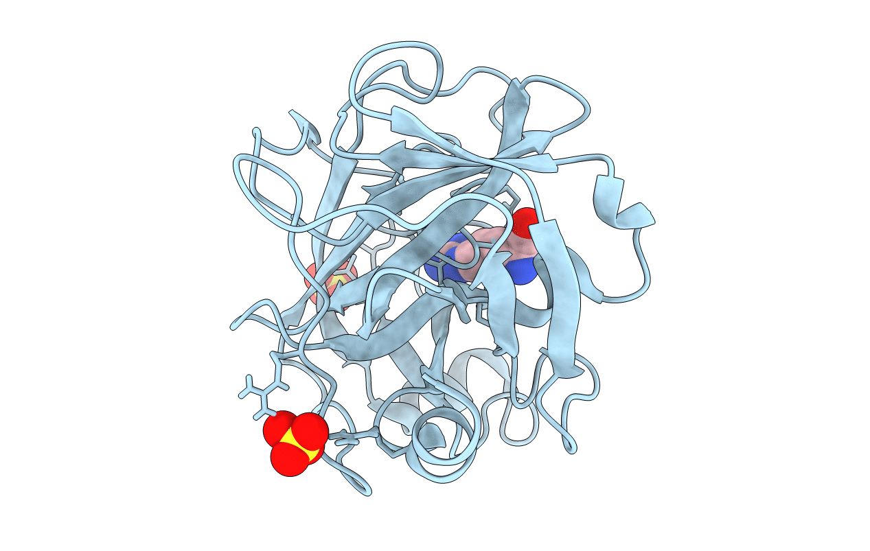
A series of crystal structures of trypsin, containing either an autoproteolytic cleaved peptide fragment or a covalently bound inhibitor, were determined at atomic and ultra-high resolution and subjected to ab initio quantum chemical calculations and multipole refinement. Quantum chemical calculations reproduced the observed active site crystal structure with severe deviations from standard stereochemistry and indicated the protonation state of the catalytic residues. Multipole refinement directly revealed the charge distribution in the active site and proved the validity of the ab initio calculations. The combined results confirmed the catalytic function of the active site residues and the two water molecules acting as the nucleophile and the proton donor. The crystal structures represent snapshots from the reaction pathway, close to a tetrahedral intermediate. The de-acylation of trypsin then occurs in true SN2 fashion.
Legend
Protein
Chemical
Disease