1PPF image
Deposition Date 1991-10-24
Release Date 1994-01-31
Last Version Date 2024-11-06
Entry Detail
PDB ID:
1PPF
Title:
X-RAY CRYSTAL STRUCTURE OF THE COMPLEX OF HUMAN LEUKOCYTE ELASTASE (PMN ELASTASE) AND THE THIRD DOMAIN OF THE TURKEY OVOMUCOID INHIBITOR
Biological Source:
Source Organism:
Homo sapiens (Taxon ID: 9606)
Meleagris gallopavo (Taxon ID: 9103)
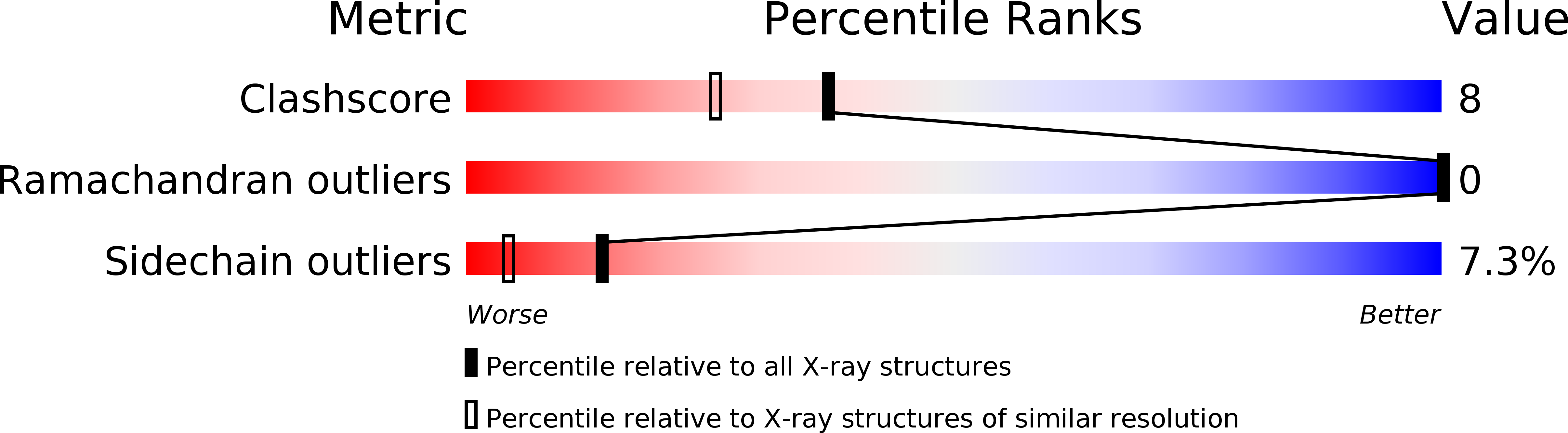
Method Details:
Experimental Method:
Resolution:
1.80 Å
R-Value Work:
0.16
Space Group:
P 21 21 21
Macromolecular Entities
Structures with similar UniProt ID
Protein Blast
Polymer Type:polypeptide(L)
Molecule:HUMAN LEUKOCYTE ELASTASE
Gene (Uniprot):ELANE
Chain IDs:A (auth: E)
Chain Length:218
Number of Molecules:1
Biological Source:Homo sapiens
Structures with similar UniProt ID
Protein Blast
Polymer Type:polypeptide(L)
Molecule:TURKEY OVOMUCOID INHIBITOR (OMTKY3)
Chain IDs:B (auth: I)
Chain Length:56
Number of Molecules:1
Biological Source:Meleagris gallopavo
Modified Residue
Compound ID Chain ID Parent Comp ID Details 2D Image
ASN A ASN GLYCOSYLATION SITE
Primary Citation
X-ray crystal structure of the complex of human leukocyte elastase (PMN elastase) and the third domain of the turkey ovomucoid inhibitor.
EMBO J. 5 2453 2458 (1986)
PMID: 3640709

Abstact

Orthorhombic crystals diffracting beyond 1.7 A resolution, have been grown from the stoichiometric complex formed between human leukocyte elastase (HLE) and the third domain of turkey ovomucoid inhibitor (OMTKY3). The crystal and molecular structure has been determined with the multiple isomorphous replacement technique. The complex has been modeled using the known structure of OMTKY3 and partial sequence information for HLE, and has been refined. The current crystallographic R-value is 0.21 for reflections from 25 to 1.8 A resolution. HLE shows the characteristic polypeptide fold of trypsin-like serine proteinases and consists of 218 amino acid residues. However, several loop segments, mainly arranged around the substrate binding site, have unique conformations. The largest deviations from the other vertebrate proteinases of known spatial structure are around Cys168. The specificity pocket is constricted by Val190, Val216 and Asp226 to preferentially accommodate medium sized hydrophobic amino acids at P1. Seven residues of the OMTKY3-binding segment are in specific contact with HLE. This interaction and geometry around the reactive site are similar as observed in other complexes. It is the first serine proteinase glycoprotein analysed, having two sugar chains attached to Asn159 and to residue 109.

Legend

Protein

Chemical

Disease

Primary Citation of related structures