Deposition Date
1994-08-05
Release Date
1995-02-07
Last Version Date
2024-02-14
Entry Detail
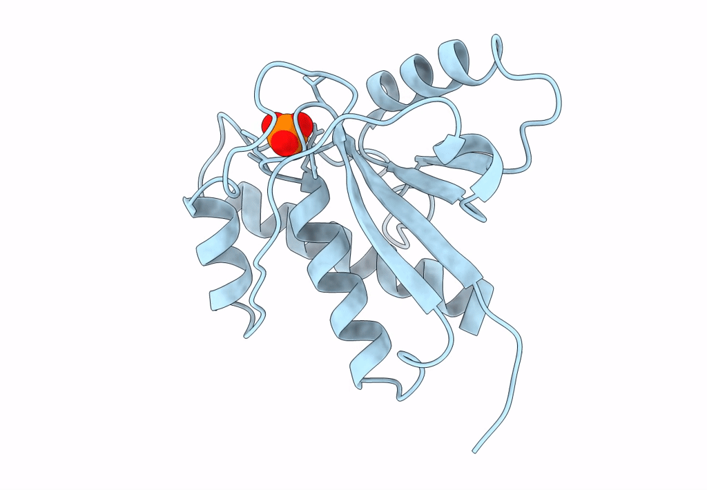
PDB ID:
1PNT
Keywords:
Title:
CRYSTAL STRUCTURE OF BOVINE HEART PHOSPHOTYROSYL PHOSPHATASE AT 2.2 ANGSTROMS RESOLUTION
Biological Source:
Source Organism(s):
Bos taurus (Taxon ID: 9913)
Method Details:
Experimental Method:
Resolution:
2.20 Å
R-Value Work:
0.16
R-Value Observed:
0.16
Space Group:
C 1 2 1