
Deposition Date
2003-06-12
Release Date
2004-04-13
Last Version Date
2023-08-16
Entry Detail
PDB ID:
1PN4
Keywords:
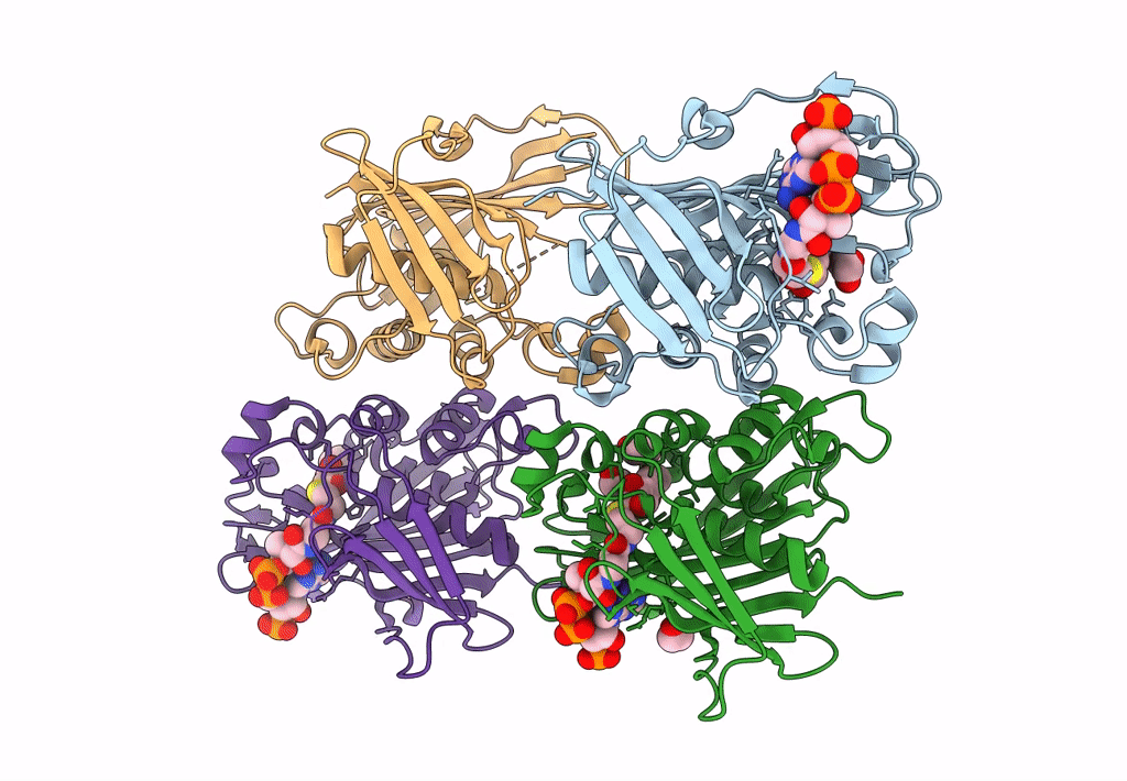
Title:
Crystal structure of 2-enoyl-CoA hydratase 2 domain of Candida tropicalis multifunctional enzyme type 2 complexed with (3R)-hydroxydecanoyl-CoA.
Biological Source:
Source Organism(s):
Candida tropicalis (Taxon ID: 5482)
Expression System(s):
Method Details:
Experimental Method:
Resolution:
2.35 Å
R-Value Free:
0.22
R-Value Work:
0.17
R-Value Observed:
0.17
Space Group:
P 1 21 1
