Deposition Date
1994-01-28
Release Date
1994-07-31
Last Version Date
2024-02-14
Entry Detail
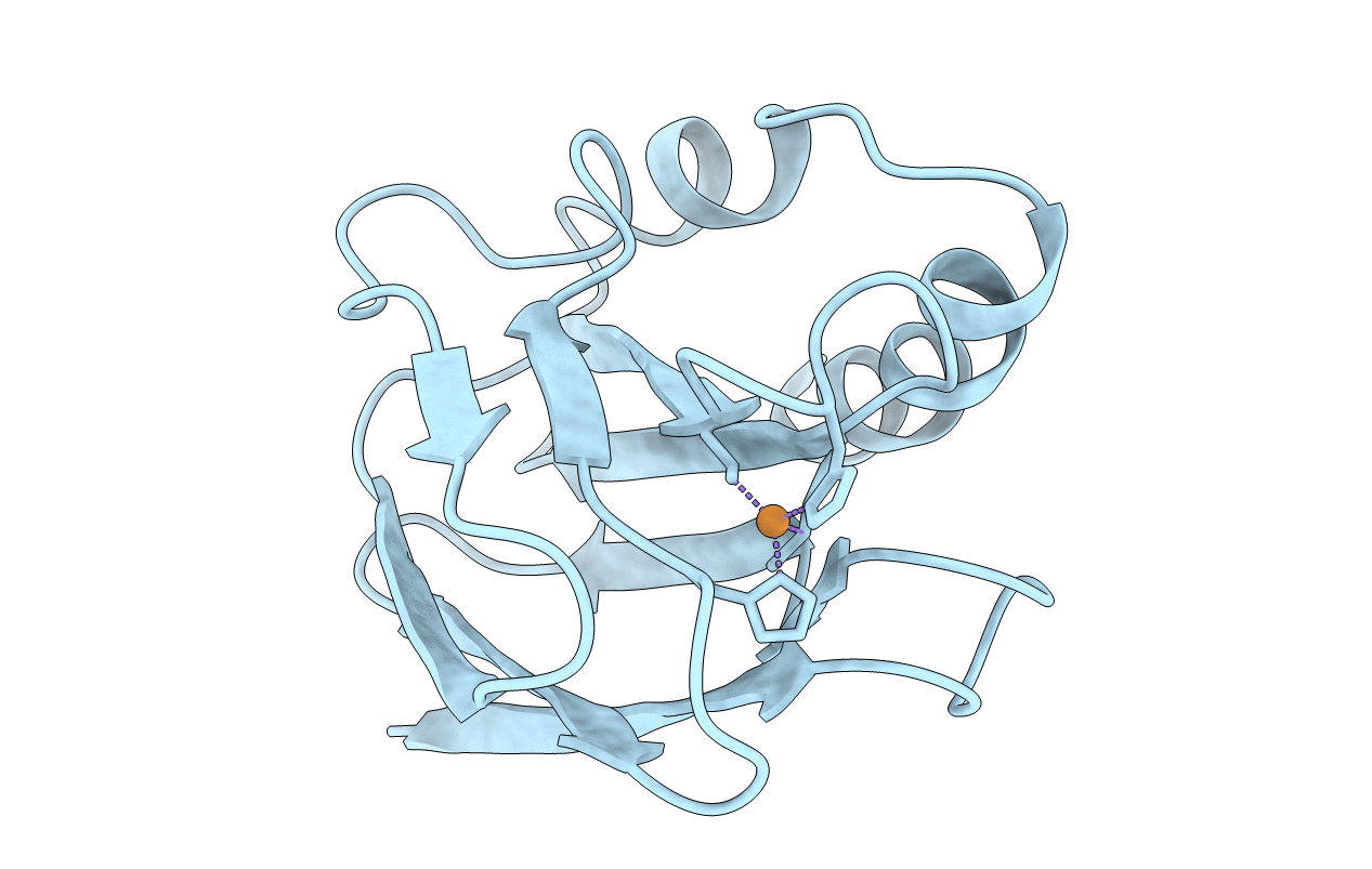
PDB ID:
1PMY
Keywords:
Title:
REFINED CRYSTAL STRUCTURE OF PSEUDOAZURIN FROM METHYLOBACTERIUM EXTORQUENS AM1 AT 1.5 ANGSTROMS RESOLUTION
Biological Source:
Source Organism(s):
Methylobacterium extorquens (Taxon ID: 408)
Method Details:
Experimental Method:
Resolution:
1.50 Å
R-Value Work:
0.19
R-Value Observed:
0.19
Space Group:
P 21 21 21