1PMO image
Deposition Date 2003-06-11
Release Date 2004-02-17
Last Version Date 2024-02-14
Entry Detail
PDB ID:
1PMO
Keywords:
Title:
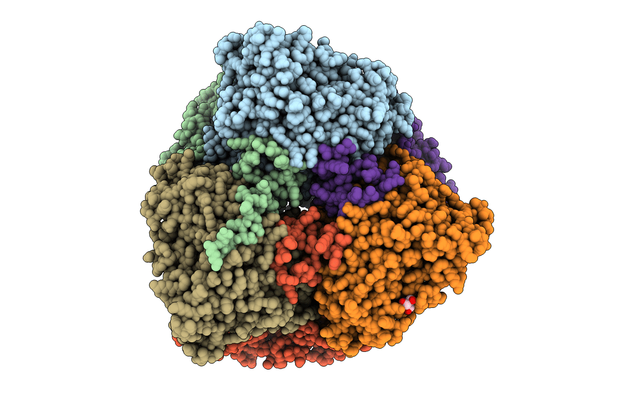
Crystal structure of Escherichia coli GadB (neutral pH)
Biological Source:
Source Organism:
Escherichia coli (Taxon ID: 562)
Host Organism:
Method Details:
Experimental Method:
Resolution:
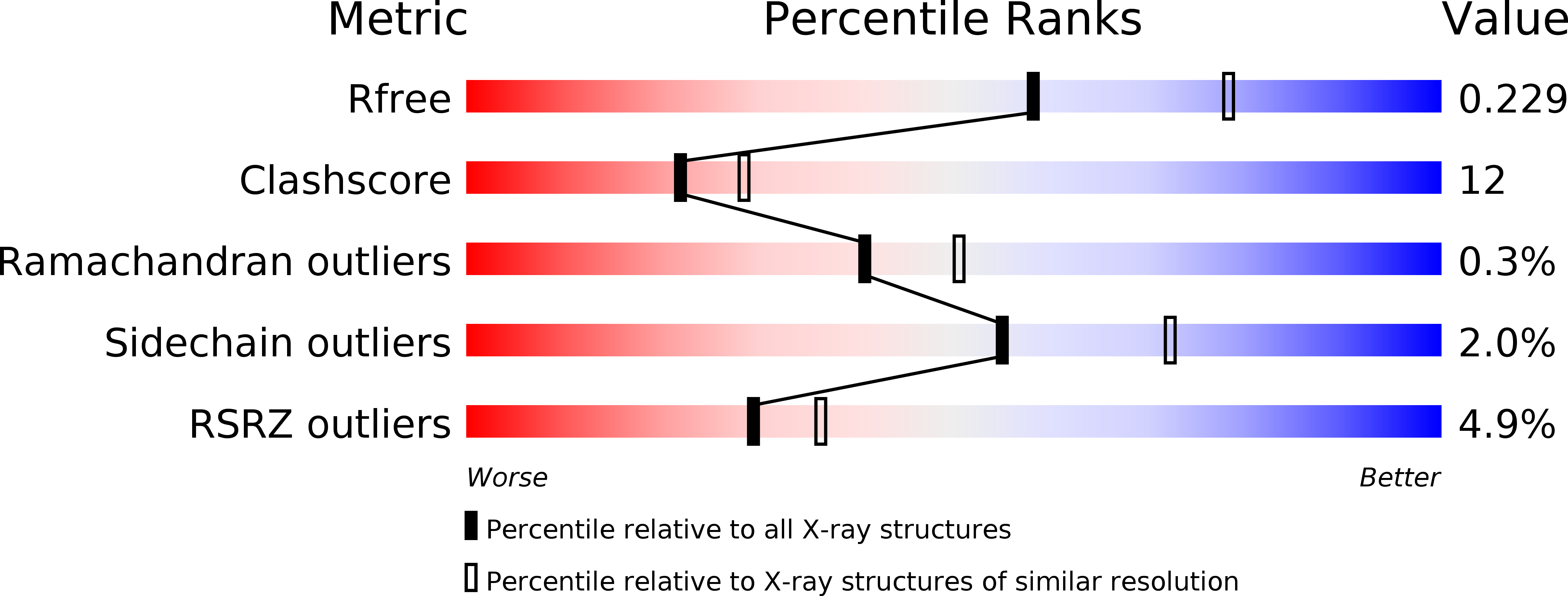
2.30 Å
R-Value Free:
0.23
R-Value Work:
0.2
Space Group:
P 32
Macromolecular Entities
Structures with similar UniProt ID
Protein Blast
Polymer Type:polypeptide(L)
Molecule:Glutamate decarboxylase beta
Gene (Uniprot):gadB
Chain IDs:A, B, C, D, E, F
Chain Length:466
Number of Molecules:6
Biological Source:Escherichia coli
Primary Citation
Crystal structure and functional analysis of escherichia coli glutamate decarboxylase
Embo J. 22 4027 4037 (2003)
PMID: 12912902 DOI: 10.1093/emboj/cdg403

Abstact

Glutamate decarboxylase is a vitamin B6-dependent enzyme, which catalyses the decarboxylation of glutamate to gamma-aminobutyrate. In Escherichia coli, expression of glutamate decarboxylase (GadB), a 330 kDa hexamer, is induced to maintain the physiological pH under acidic conditions, like those of the passage through the stomach en route to the intestine. GadB, together with the antiporter GadC, constitutes the gad acid resistance system, which confers the ability for bacterial survival for at least 2 h in a strongly acidic environment. GadB undergoes a pH-dependent conformational change and exhibits an activity optimum at low pH. We determined the crystal structures of GadB at acidic and neutral pH. They reveal the molecular details of the conformational change and the structural basis for the acidic pH optimum. We demonstrate that the enzyme is localized exclusively in the cytoplasm at neutral pH, but is recruited to the membrane when the pH falls. We show by structure-based site-directed mutagenesis that the triple helix bundle formed by the N-termini of the protein at acidic pH is the major determinant for this behaviour.

Legend

Protein

Chemical

Disease

Primary Citation of related structures