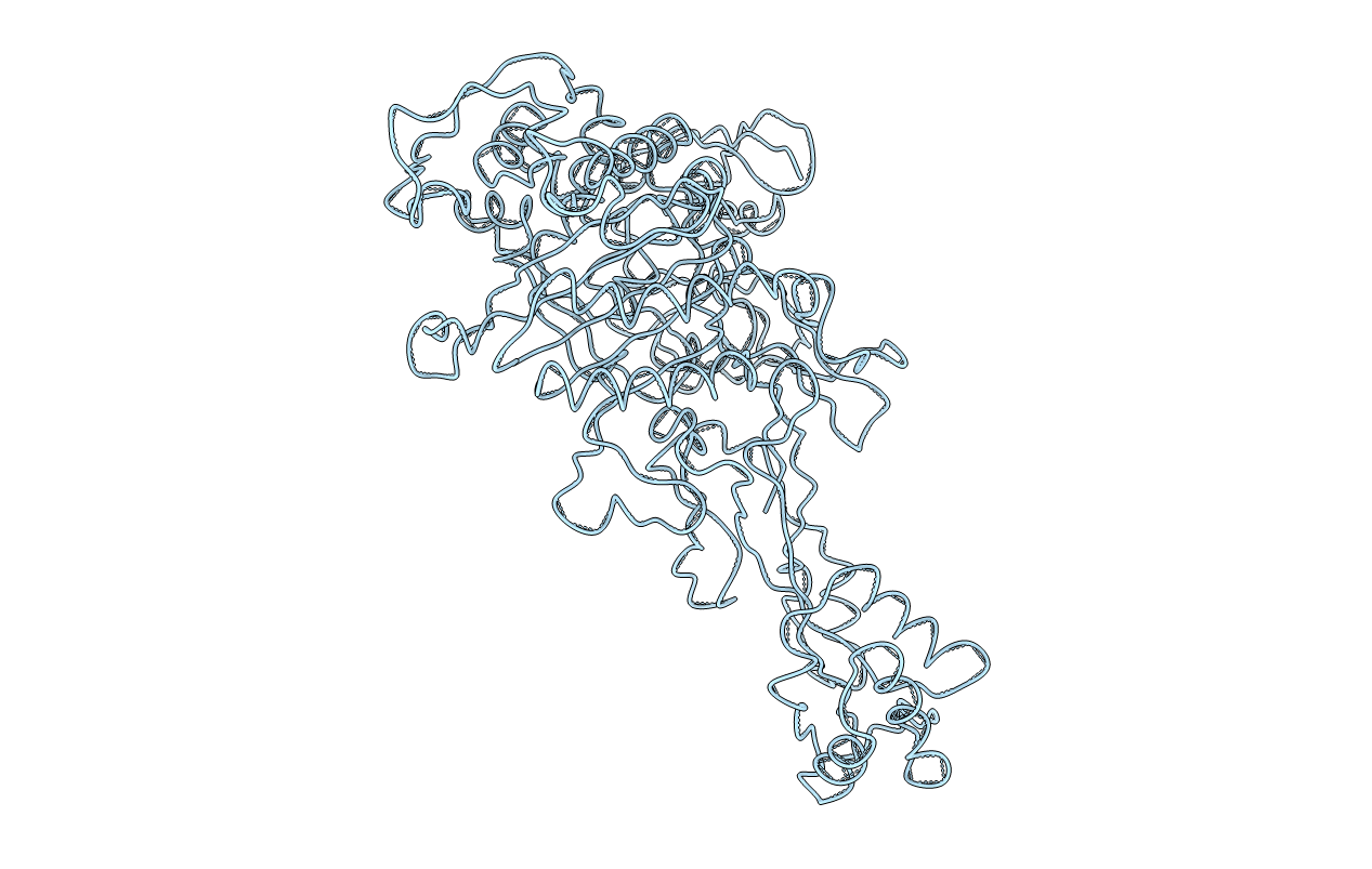
1PMD image
Entry Detail
PDB ID:
1PMD
Title:
PENICILLIN-BINDING PROTEIN 2X (PBP-2X)
Biological Source:
Source Organism:
PDB Version:
Deposition Date:
1996-02-05
Release Date:
1997-02-05
Method Details:
Experimental Method:
Resolution:
3.50 Å
R-Value Free:
0.21
R-Value Work:
0.22
R-Value Observed:
0.22
Space Group:
P 61 2 2
Macromolecular Entities
Polymer Type:polypeptide(L)
Description:PEPTIDOGLYCAN SYNTHESIS MULTIFUNCTIONAL ENZYME
Chain IDs:A
Chain Length:675
Number of Molecules:1
Biological Source:Streptococcus pneumoniae
Ligand Molecules
Primary Citation
X-ray structure of Streptococcus pneumoniae PBP2x, a primary penicillin target enzyme.
Nat.Struct.Biol. 3 284 289 (1996)
PMID: 8605631 DOI: 10.1038/nsb0396-284

Abstact

All beta-lactam antibiotics exert their biological effects by interacting with a unique class of proteins, the penicillin-binding proteins (PBPs). These membrane proteins are involved in the biosynthesis of the murein or peptidoglycan, a mesh-like structure which completely surrounds the bacterial cell. Sequence similarities indicate that one domain of these proteins belongs to a large family of beta-lactam-recognizing proteins, which includes the active-site serine beta-lactamases. We here report the first three-dimensional crystal structure of a high molecular weight penicillin-binding protein, PBP2x of Streptococcus pneumoniae, at 3.5 A resolution. The molecule has three domains, the central domain being a transpeptidase, which is a suitable target for antibiotic development.

Legend

Protein

Chemical

Disease

Primary Citation of related structures