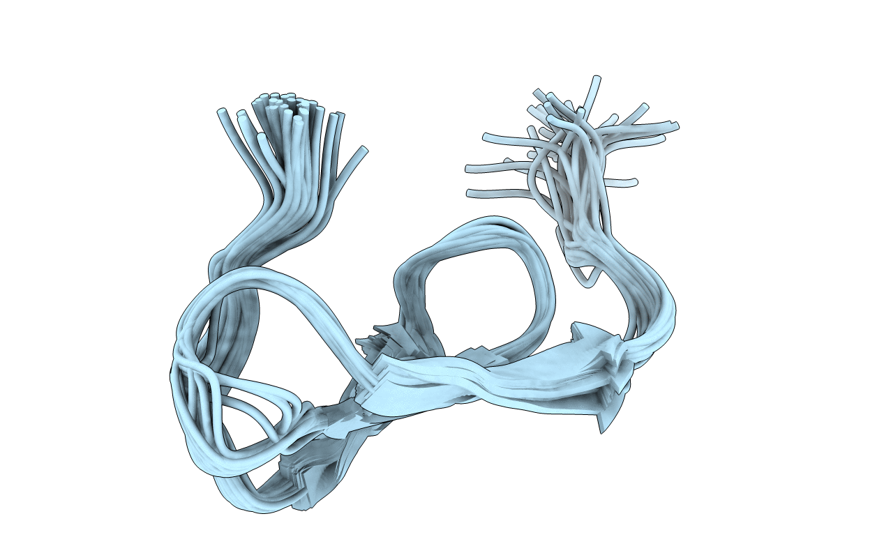
The solution structure and the disulfide pairings of a 36-residue proteinase inhibitor isolated from the insect Locusta migratoria have been determined using NMR spectroscopy and simulated annealing calculations. The peptide, termed PMP-C, was previously shown to inhibit bovine alpha-chymotrypsin as well as human leukocyte elastase, and was also found to block high-voltage-activated Ca2+ currents in rat sensory neurones. PMP-C has a prolate ellipsoid shape and adopts a tertiary fold hitherto unobserved in the large group of small "canonical" proteinase inhibitors. The over-all fold consists mainly of three strands arranged in a right-handed twisted, antiparallel, beta-sheet that demarcates a cavity, together with a linear amino-terminal segment oriented almost perpendicular to the three strands of the beta-sheet. Inside the cavity a phenyl ring constitutes the centre of a hydrophobic core. The proteinase binding loop is located in the carboxy-terminal part of the molecule, between two cysteine residues involved in disulfide bridges. Its conformation resembles that found in other small canonical proteinase inhibitors. A comparison of PMP-C structure with the recently published solution structure of the related peptide PMP-D2 shows that the most significant differences are complementary changes involved in the stabilization of similar folds. This comparison led us to review the structure of PMP-D2 and to identify two salt bridges in PMP-D2.
Legend
Protein
Chemical
Disease