Deposition Date
2003-06-09
Release Date
2004-01-27
Last Version Date
2024-11-06
Entry Detail
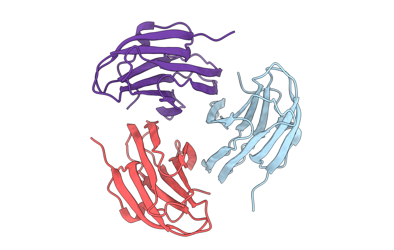
PDB ID:
1PM4
Keywords:
Title:
Crystal structure of Yersinia pseudotuberculosis-derived mitogen (YPM)
Biological Source:
Source Organism(s):
Yersinia pseudotuberculosis (Taxon ID: 633)
Expression System(s):
Method Details:
Experimental Method:
Resolution:
1.76 Å
R-Value Free:
0.22
R-Value Work:
0.18
R-Value Observed:
0.18
Space Group:
C 1 2 1