1PL3 image
Deposition Date 2003-06-06
Release Date 2003-07-01
Last Version Date 2024-11-13
Entry Detail
PDB ID:
1PL3
Keywords:
Title:
Cytochrome Domain Of Cellobiose Dehydrogenase, M65H mutant
Biological Source:
Source Organism:
Method Details:
Experimental Method:
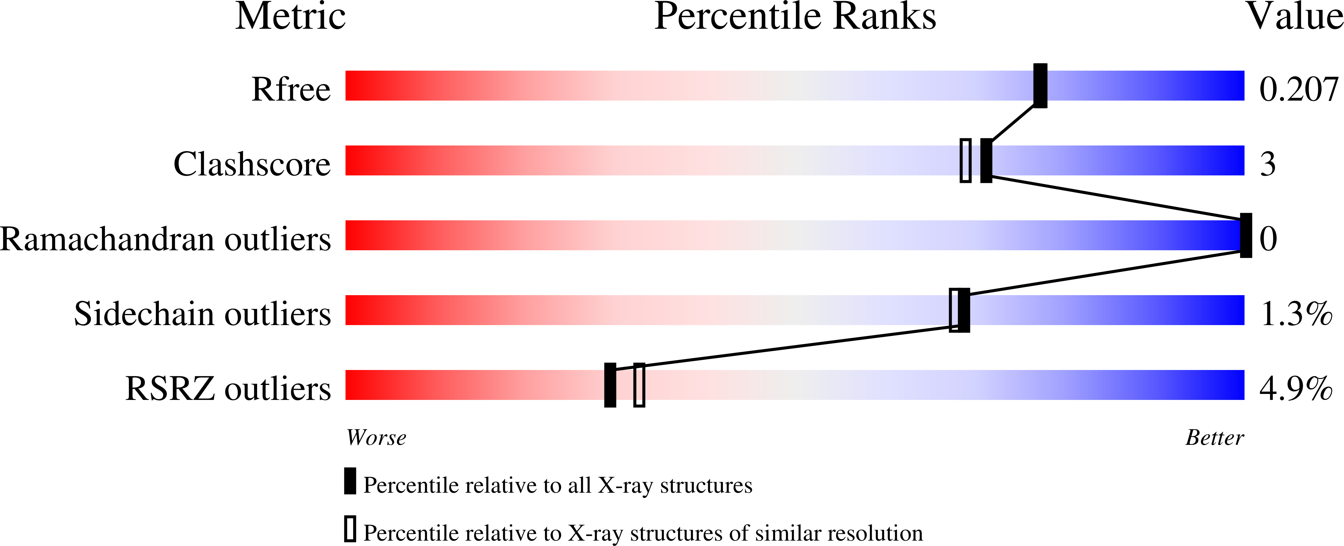
Resolution:
1.90 Å
R-Value Free:
0.19
R-Value Work:
0.17
R-Value Observed:
0.17
Space Group:
P 65
Macromolecular Entities
Polymer Type:polypeptide(L)
Molecule:Cellobiose dehydrogenase
Gene (Uniprot):CDH-1, CDH-2
Mutations:M65H
Chain IDs:A, B
Chain Length:186
Number of Molecules:2
Biological Source:Phanerochaete chrysosporium
Modified Residue
Compound ID Chain ID Parent Comp ID Details 2D Image
ASN A ASN GLYCOSYLATION SITE
PCA A GLN PYROGLUTAMIC ACID
Primary Citation
Biophysical and Structural Analysis of a Novel Heme b Iron Ligation in the Flavocytochrome Cellobiose Dehydrogenase.
J.Biol.Chem. 278 33224 33231 (2003)
PMID: 12796496 DOI: 10.1074/jbc.M302653200

Abstact

The fungal extracellular flavocytochrome cellobiose dehydrogenase (CDH) participates in lignocellulose degradation. The enzyme has a cytochrome domain connected to a flavin-binding domain by a peptide linker. The cytochrome domain contains a 6-coordinate low spin b-type heme with unusual iron ligands and coordination geometry. Wild type CDH is only the second example of a b-type heme with Met-His ligation, and it is the first example of a Met-His ligation of heme b where the ligands are arranged in a nearly perpendicular orientation. To investigate the ligation further, Met65 was replaced with a histidine to create a bis-histidyl ligated iron typical of b-type cytochromes. The variant is expressed as a stable 90-kDa protein that retains the flavin domain catalytic reactivity. However, the ability of the mutant to reduce external one-electron acceptors such as cytochrome c is impaired. Electrochemical measurements demonstrate a decrease in the redox midpoint potential of the heme by 210 mV. In contrast to the wild type enzyme, the ferric state of the protoheme displays a mixed low spin/high spin state at room temperature and low spin character at 90 K, as determined by resonance Raman spectroscopy. The wild type cytochrome does not bind CO, but the ferrous state of the variant forms a CO complex, although the association rate is very low. The crystal structure of the M65H cytochrome domain has been determined at 1.9 A resolution. The variant structure confirms a bis-histidyl ligation but reveals unusual features. As for the wild type enzyme, the ligands have a nearly perpendicular arrangement. Furthermore, the iron is bound by imidazole N delta 1 and N epsilon 2 nitrogen atoms, rather than the typical N epsilon 2/N epsilon 2 coordination encountered in bis-histidyl ligated heme proteins. To our knowledge, this is the first example of a bis-histidyl N delta 1/N epsilon 2-coordinated protoporphyrin IX iron.

Legend

Protein

Chemical

Disease

Primary Citation of related structures