1PKV image
Deposition Date 2003-06-06
Release Date 2004-06-08
Last Version Date 2023-08-16
Entry Detail
PDB ID:
1PKV
Keywords:
Title:
The N-terminal domain of riboflavin synthase in complex with riboflavin
Biological Source:
Source Organism:
Escherichia coli (Taxon ID: 562)
Host Organism:
Method Details:
Experimental Method:
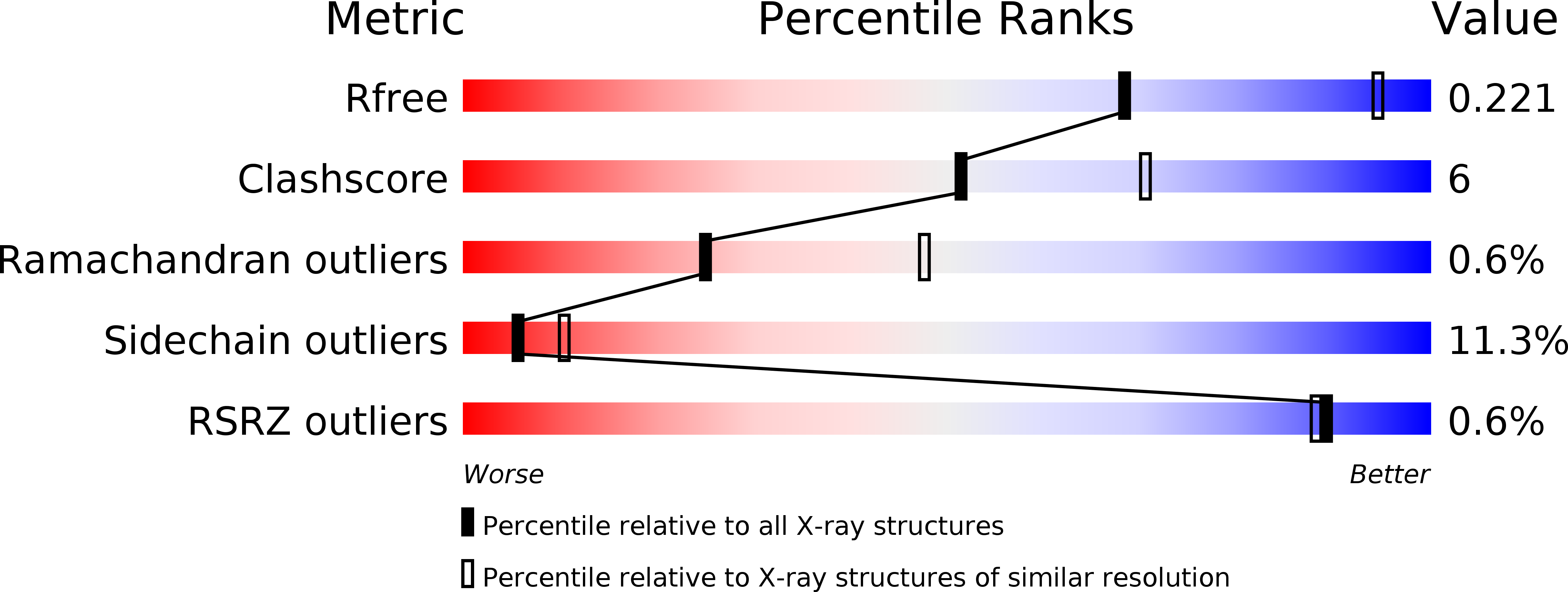
Resolution:
2.60 Å
R-Value Free:
0.23
R-Value Work:
0.17
R-Value Observed:
0.17
Space Group:
C 2 2 21
Macromolecular Entities
Polymer Type:polypeptide(L)
Molecule:Riboflavin synthase alpha chain
Chain IDs:A, B
Chain Length:97
Number of Molecules:2
Biological Source:Escherichia coli
Ligand Molecules
Primary Citation
The structure of the N-terminal domain of riboflavin synthase in complex with riboflavin at 2.6A resolution.
J.Mol.Biol. 331 1053 1063 (2003)
PMID: 12927541 DOI: 10.1016/S0022-2836(03)00844-1

Abstact

Riboflavin synthase of Escherichia coli is a homotrimer with a molecular mass of 70 kDa. The enzyme catalyzes the dismutation of 6,7-dimethyl-8-(1'-D-ribityl)-lumazine, affording riboflavin and 5-amino-6-ribitylamino-2,4(1H,3H)-pyrimidinedione. The N-terminal segment (residues 1-87) and the C-terminal segment (residues 98-187) form beta-barrels with similar fold and a high degree of sequence similarity. A recombinant peptide comprising amino acid residues 1-97 forms a dimer, which binds riboflavin with high affinity. Here, we report the structure of this construct in complex with riboflavin at 2.6A resolution. It is demonstrated that the complex can serve as a model for ligand-binding in the native enzyme. The structure and riboflavin-binding mode is in excellent agreement with structural information obtained from the native enzyme from Escherichia coli and riboflavin synthase from Schizosaccharomyces pombe. The implications for the binding specificity and the regiospecificity of the catalyzed reaction are discussed.

Legend

Protein

Chemical

Disease

Primary Citation of related structures