
Deposition Date
1994-03-07
Release Date
1994-05-31
Last Version Date
2024-05-01
Entry Detail
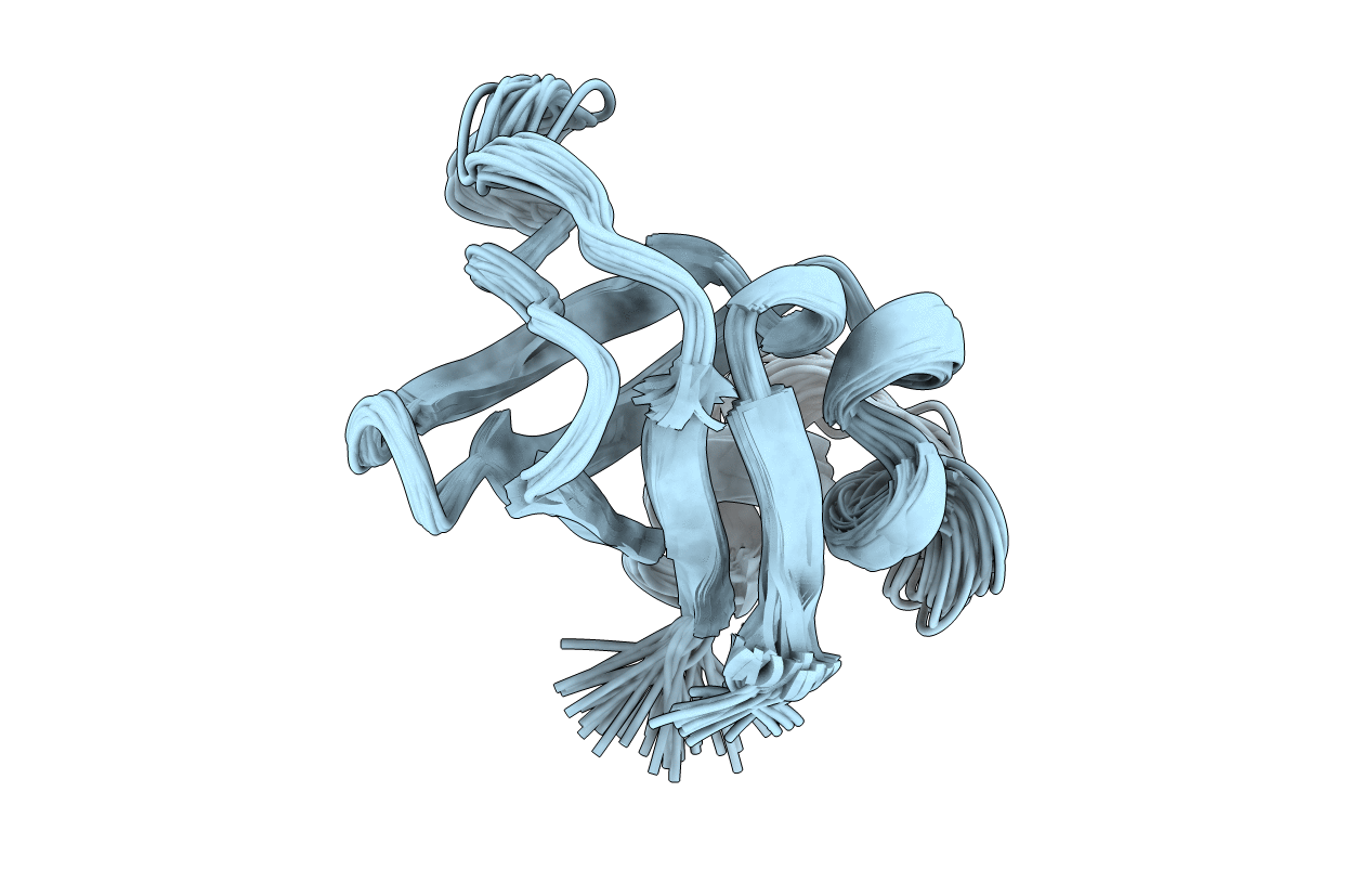
PDB ID:
1PKT
Keywords:
Title:
STRUCTURE OF THE PI3K SH3 DOMAIN AND ANALYSIS OF THE SH3 FAMILY
Biological Source:
Source Organism(s):
Homo sapiens (Taxon ID: 9606)
Method Details:
Experimental Method:
Conformers Submitted:
30
