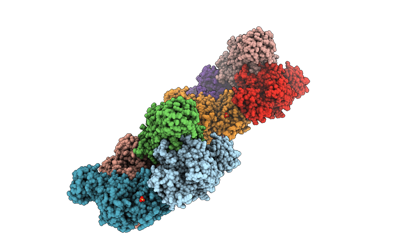
1PKL image
Deposition Date 1998-09-15
Release Date 1998-09-23
Last Version Date 2023-08-16
Entry Detail
PDB ID:
1PKL
Keywords:
Title:
THE STRUCTURE OF LEISHMANIA PYRUVATE KINASE
Biological Source:
Source Organism:
Host Organism:
Method Details:
Experimental Method:
Resolution:
2.35 Å
R-Value Free:
0.25
R-Value Work:
0.20
R-Value Observed:
0.20
Space Group:
P 1 21 1
Macromolecular Entities
Structures with similar UniProt ID
Protein Blast
Polymer Type:polypeptide(L)
Molecule:PROTEIN (PYRUVATE KINASE)
Gene (Uniprot):PYK
Chain IDs:A, B, C, D, E, F, G (auth: H), H (auth: G)
Chain Length:499
Number of Molecules:8
Biological Source:Leishmania mexicana
Ligand Molecules
Primary Citation
The structure of pyruvate kinase from Leishmania mexicana reveals details of the allosteric transition and unusual effector specificity.
J.Mol.Biol. 291 615 635 (1999)
PMID: 10448041 DOI: 10.1006/jmbi.1999.2918

Abstact

Glycolysis occupies a central role in cellular metabolism, and is of particular importance for the catabolic production of ATP in protozoan parasites such as Leishmania and Trypanosoma. In these organisms pyruvate kinase plays a key regulatory role, and is unique in responding to fructose 2,6-bisphosphate as allosteric activator. The determination of the first eukaryotic pyruvate kinase crystal structure in the T-state is reported. A comparison of the leishmania and yeast R-state enzymes reveals fewer differences than the previous comparison of Escherichia coli T-state and rabbit muscle non-allosteric enzymes. Structural changes related to the allosteric transition can therefore be distinguished from those that are a consequence of the inherent wide structural divergence between bacterial and mammalian proteins. The allosteric transition involves significant changes in a tightly packed array of eight alpha helices at the interface near the catalytic site. At the other interface the allosteric transition appears to be accompanied by the bending of a ten-stranded intersubunit beta sheet adjacent to the effector site. Helix Calpha1 makes contacts to the N-terminal helical domain and bridges both interfaces. A comparison of the effector sites of the leishmania and yeast enzymes reveals the structural basis for the different effector specificity. Two loops comprising residues 443-453 and 480-489 adopt very different conformations in the two enzymes, and Lys453 and His480 that are a feature of trypanosomatid enzymes provide probable ligands for the 2-phospho group of the effector molecule. These differences offer an opportunity for the design of drugs that would bind to the trypanosomatid enzymes but not to those of the mammalian host.

Legend

Protein

Chemical

Disease

Primary Citation of related structures