1PJP image
Entry Detail
PDB ID:
1PJP
Title:
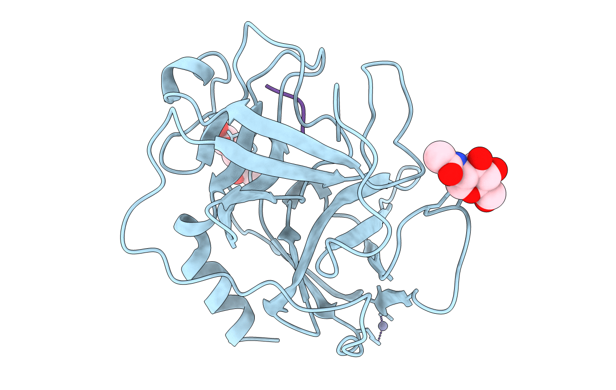
THE 2.2 A CRYSTAL STRUCTURE OF HUMAN CHYMASE IN COMPLEX WITH SUCCINYL-ALA-ALA-PRO-PHE-CHLOROMETHYLKETONE
Biological Source:
Host Organism:
PDB Version:
Deposition Date:
1998-09-07
Release Date:
1999-03-02
Method Details:
Experimental Method:
Resolution:
2.20 Å
R-Value Free:
0.23
R-Value Work:
0.18
R-Value Observed:
0.18
Space Group:
P 43
Macromolecular Entities
Polymer Type:polypeptide(L)
Description:Chymase
Mutations:F127K, V208A, R235Q
Chain IDs:A
Chain Length:226
Number of Molecules:1
Biological Source:Homo sapiens
Polymer Type:polypeptide(L)
Description:SUCCINYL-ALA-ALA-PRO-PHE-CHLOROMETHYLKETONE INHIBITOR
Chain IDs:B (auth: I)
Chain Length:5
Number of Molecules:1
Biological Source:synthetic construct
Modified Residue
Compound ID Chain ID Parent Comp ID Details 2D Image
ASN A ASN GLYCOSYLATION SITE
HPH B PHE (2S)-2-AMINO-3-PHENYLPROPANE-1,1-DIOL
Peptide-like Molecules
PRD_000381
Primary Citation
The 2.2 A crystal structure of human chymase in complex with succinyl-Ala-Ala-Pro-Phe-chloromethylketone: structural explanation for its dipeptidyl carboxypeptidase specificity.
J.Mol.Biol. 286 163 173 (1999)
PMID: 9931257 DOI: 10.1006/jmbi.1998.2462

Abstact

Human chymase (HC) is a chymotrypsin-like serine proteinase expressed by mast cells. The 2.2 A crystal structure of HC complexed to the peptidyl inhibitor, succinyl-Ala-Ala-Pro-Phe-chloromethylketone (CMK), was solved and refined to a crystallographic R-factor of 18.4 %. The HC structure exhibits the typical folding pattern of a chymotrypsin-like serine proteinase, and shows particularly similarity to rat chymase 2 (rat mast cell proteinase II) and human cathepsin G. The peptidyl-CMK inhibitor is covalently bound to the active-site residues Ser195 and His57; the peptidyl moiety juxtaposes the S1 entrance frame segment 214-217 by forming a short antiparallel beta-sheet. HC is a highly efficient angiotensin-converting enzyme. Modeling of the chymase-angiotensin I interaction guided by the geometry of the bound chloromethylketone inhibitor indicates that the extended substrate binding site contains features that may generate the dipeptidyl carboxypeptidase-like activity needed for efficient cleavage and activation of the hormone. The C-terminal carboxylate group of angiotensin I docked into the active-site cleft, with the last two residues extending beyond the active site, is perfectly localized to make a favorable hydrogen bond and salt bridge with the amide nitrogen of the Lys40-Phe41 peptide bond and with the epsilon-ammonium group of the Lys40 side-chain. This amide positioning is unique to the chymase-related proteinases, and only chymases from primates possess a Lys residue at position 40. Thus, the structure conveniently explains the preferred conversion of angiotensin I to angiotensin II by human chymase.

Legend

Protein

Chemical

Disease

Primary Citation of related structures