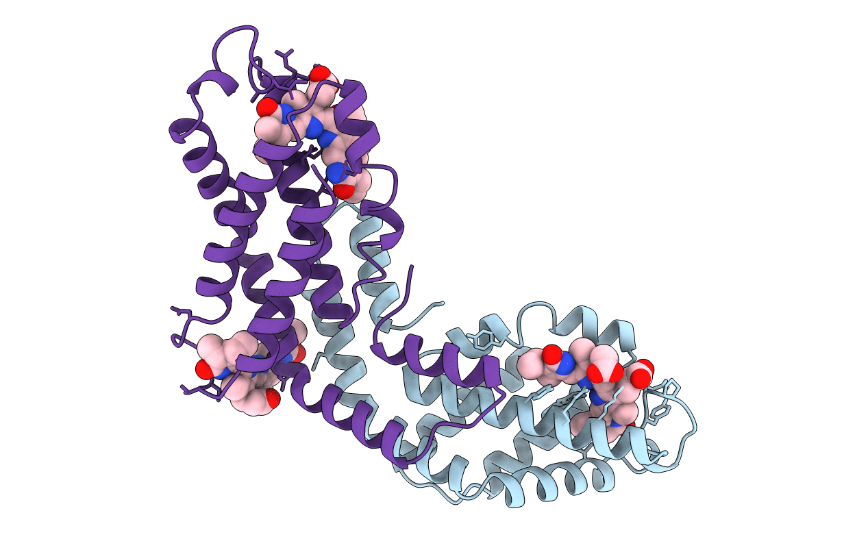
1PHN image
Entry Detail
PDB ID:
1PHN
Title:
STRUCTURE OF PHYCOCYANIN FROM CYANIDIUM CALDARIUM AT 1.65A RESOLUTION
Biological Source:
Source Organism:
PDB Version:
Deposition Date:
1995-06-21
Release Date:
1997-09-17
Method Details:
Experimental Method:
Resolution:
1.65 Å
R-Value Free:
0.27
R-Value Work:
0.18
R-Value Observed:
0.18
Space Group:
H 3 2
Macromolecular Entities
Polymer Type:polypeptide(L)
Description:PHYCOCYANIN
Chain IDs:A
Chain Length:162
Number of Molecules:1
Biological Source:Cyanidium caldarium
Polymer Type:polypeptide(L)
Description:PHYCOCYANIN
Chain IDs:B
Chain Length:172
Number of Molecules:1
Biological Source:Cyanidium caldarium
Modified Residue
Compound ID Chain ID Parent Comp ID Details 2D Image
MEN B ASN N-METHYL ASPARAGINE
Primary Citation
Crystal structure of C-phycocyanin from Cyanidium caldarium provides a new perspective on phycobilisome assembly.
Biophys.J. 76 2912 2921 (1999)
PMID: 10354419

Abstact

The crystal structure of the light-harvesting protein phycocyanin from the cyanobacterium Cyanidium caldarium with novel crystal packing has been solved at 1.65-A resolution. The structure has been refined to an R value of 18.3% with excellent backbone and side-chain stereochemical parameters. In crystals of phycocyanin used in this study, the hexamers are offset rather than aligned as in other phycocyanins that have been crystallized to date. Analysis of this crystal's unique packing leads to a proposal for phycobilisome assembly in vivo and for a more prominent role for chromophore beta-155. This new role assigned to chromophore beta-155 in phycocyanin sheds light on the numerical relationships among and function of external chromophores found in phycoerythrins and phycoerythrocyanins.

Legend

Protein

Chemical

Disease

Primary Citation of related structures