
Deposition Date
1998-03-20
Release Date
1998-05-27
Last Version Date
2024-11-20
Entry Detail
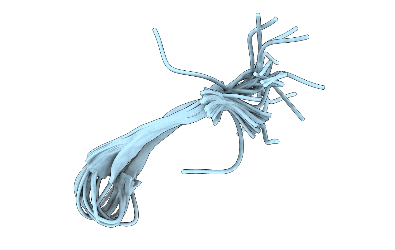
PDB ID:
1PG1
Keywords:
Title:
PROTEGRIN 1 (PG1) FROM PORCINE LEUKOCYTES, NMR, 20 STRUCTURES
Biological Source:
Source Organism(s):
Sus scrofa (Taxon ID: 9823)
Method Details:
Experimental Method:
Conformers Calculated:
100
Conformers Submitted:
20
Selection Criteria:
LEAST RESTRAINT VIOLATION, LEAST GEOMETRY VIOLATION
