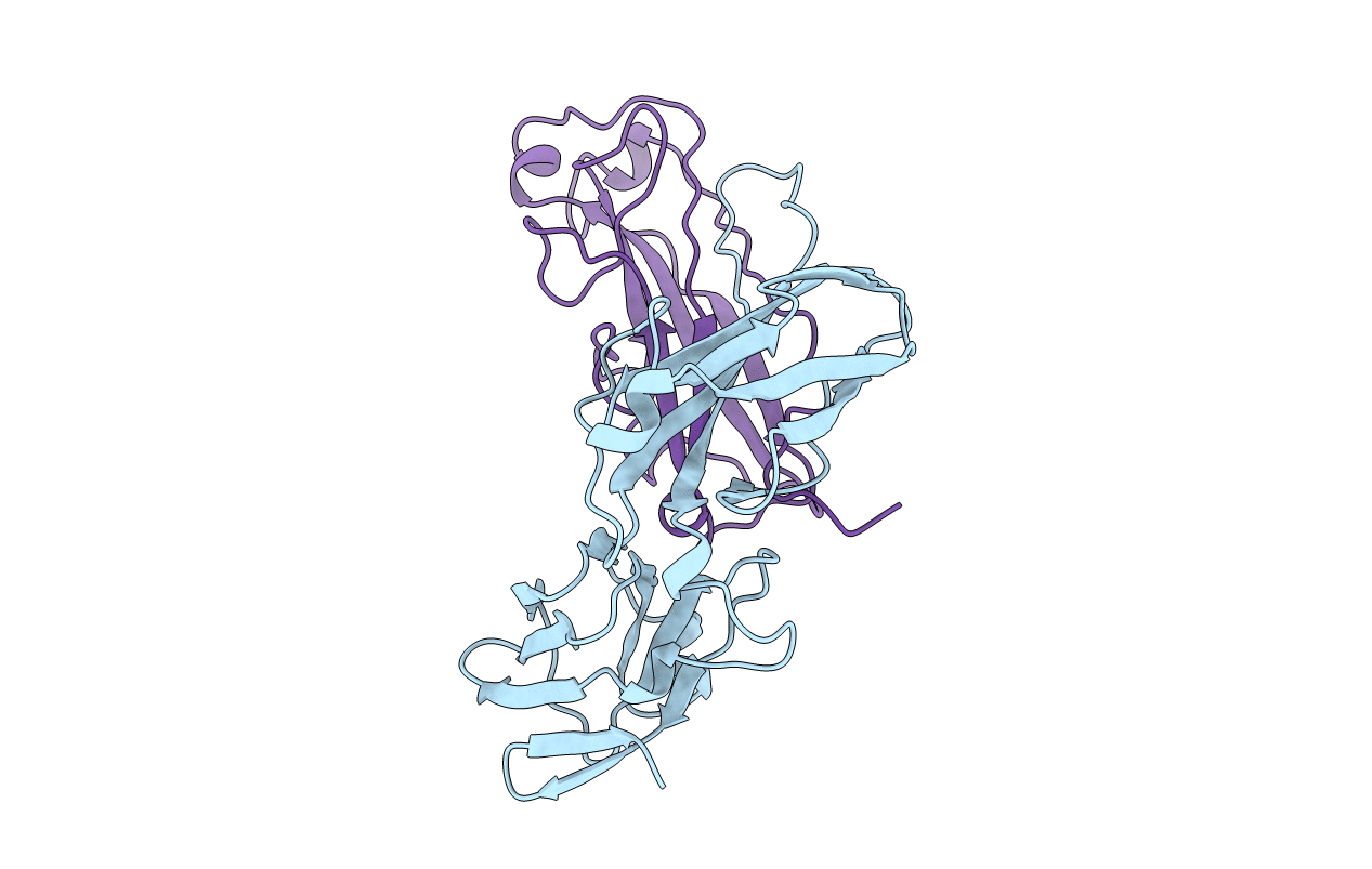
1PDK image
Deposition Date 1999-03-29
Release Date 1999-08-17
Last Version Date 2024-10-30
Entry Detail
PDB ID:
1PDK
Keywords:
Title:
PAPD-PAPK CHAPERONE-PILUS SUBUNIT COMPLEX FROM E.COLI P PILUS
Biological Source:
Source Organism:
Escherichia coli (Taxon ID: 562)
Method Details:
Experimental Method:
Resolution:
2.40 Å
R-Value Free:
0.27
R-Value Work:
0.23
Space Group:
P 21 21 21
Macromolecular Entities
Polymer Type:polypeptide(L)
Molecule:PROTEIN (CHAPERONE PROTEIN PAPD)
Gene (Uniprot):papD
Chain IDs:A
Chain Length:218
Number of Molecules:1
Biological Source:Escherichia coli
Polymer Type:polypeptide(L)
Molecule:PROTEIN (PROTEIN PAPK)
Gene (Uniprot):papK
Chain IDs:B
Chain Length:157
Number of Molecules:1
Biological Source:Escherichia coli
Primary Citation
Structural basis of chaperone function and pilus biogenesis.
Science 285 1058 1061 (1999)
PMID: 10446050 DOI: 10.1126/science.285.5430.1058

Abstact

Many Gram-negative pathogens assemble architecturally and functionally diverse adhesive pili on their surfaces by the chaperone-usher pathway. Immunoglobulin-like periplasmic chaperones escort pilus subunits to the usher, a large protein complex that facilitates the translocation and assembly of subunits across the outer membrane. The crystal structure of the PapD-PapK chaperone-subunit complex, determined at 2.4 angstrom resolution, reveals that the chaperone functions by donating its G(1) beta strand to complete the immunoglobulin-like fold of the subunit via a mechanism termed donor strand complementation. The structure of the PapD-PapK complex also suggests that during pilus biogenesis, every subunit completes the immunoglobulin-like fold of its neighboring subunit via a mechanism termed donor strand exchange.

Legend

Protein

Chemical

Disease

Primary Citation of related structures