1PD2 image
Deposition Date 1998-12-14
Release Date 1999-10-13
Last Version Date 2023-12-27
Entry Detail
PDB ID:
1PD2
Keywords:
Title:
CRYSTAL STRUCTURE OF HEMATOPOIETIC PROSTAGLANDIN D SYNTHASE COMPLEX WITH GLUTATHIONE
Biological Source:
Source Organism:
Method Details:
Experimental Method:
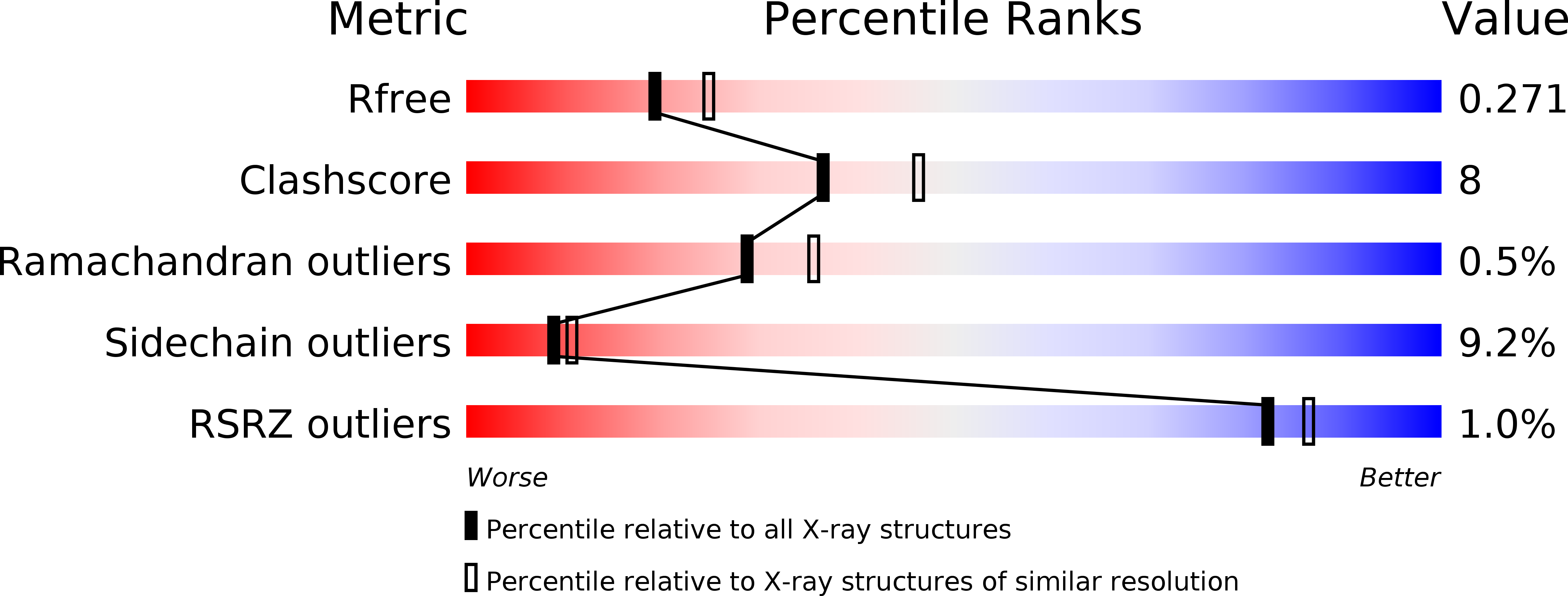
Resolution:
2.30 Å
R-Value Free:
0.28
R-Value Work:
0.20
Space Group:
P 31 2 1
Macromolecular Entities
Polymer Type:polypeptide(L)
Molecule:HEMATOPOIETIC PROSTAGLANDIN D SYNTHASE
Gene (Uniprot):Hpgds
Chain IDs:A (auth: 1), B (auth: 2)
Chain Length:199
Number of Molecules:2
Biological Source:Rattus norvegicus
Ligand Molecules
Primary Citation
Cloning and crystal structure of hematopoietic prostaglandin D synthase.
Cell(Cambridge,Mass.) 90 1085 1095 (1997)
PMID: 9323136 DOI: 10.1016/S0092-8674(00)80374-8

Abstact

Hematopoietic prostaglandin (PG) D synthase is the key enzyme for production of the D and J series of prostanoids in the immune system and mast cells. We isolated a cDNA for the rat enzyme, crystallized the recombinant enzyme, and determined the three-dimensional structure of the enzyme complexed with glutathione at 2.3 A resolution. The enzyme is the first member of the sigma class glutathione S-transferase (GST) from vertebrates and possesses a prominent cleft as the active site, which is never seen among other members of the GST family. The unique 3-D architecture of the cleft leads to the putative substrate binding mode and its catalytic mechanism, responsible for the specific isomerization from PGH2 to PGD2.

Legend

Protein

Chemical

Disease

Primary Citation of related structures