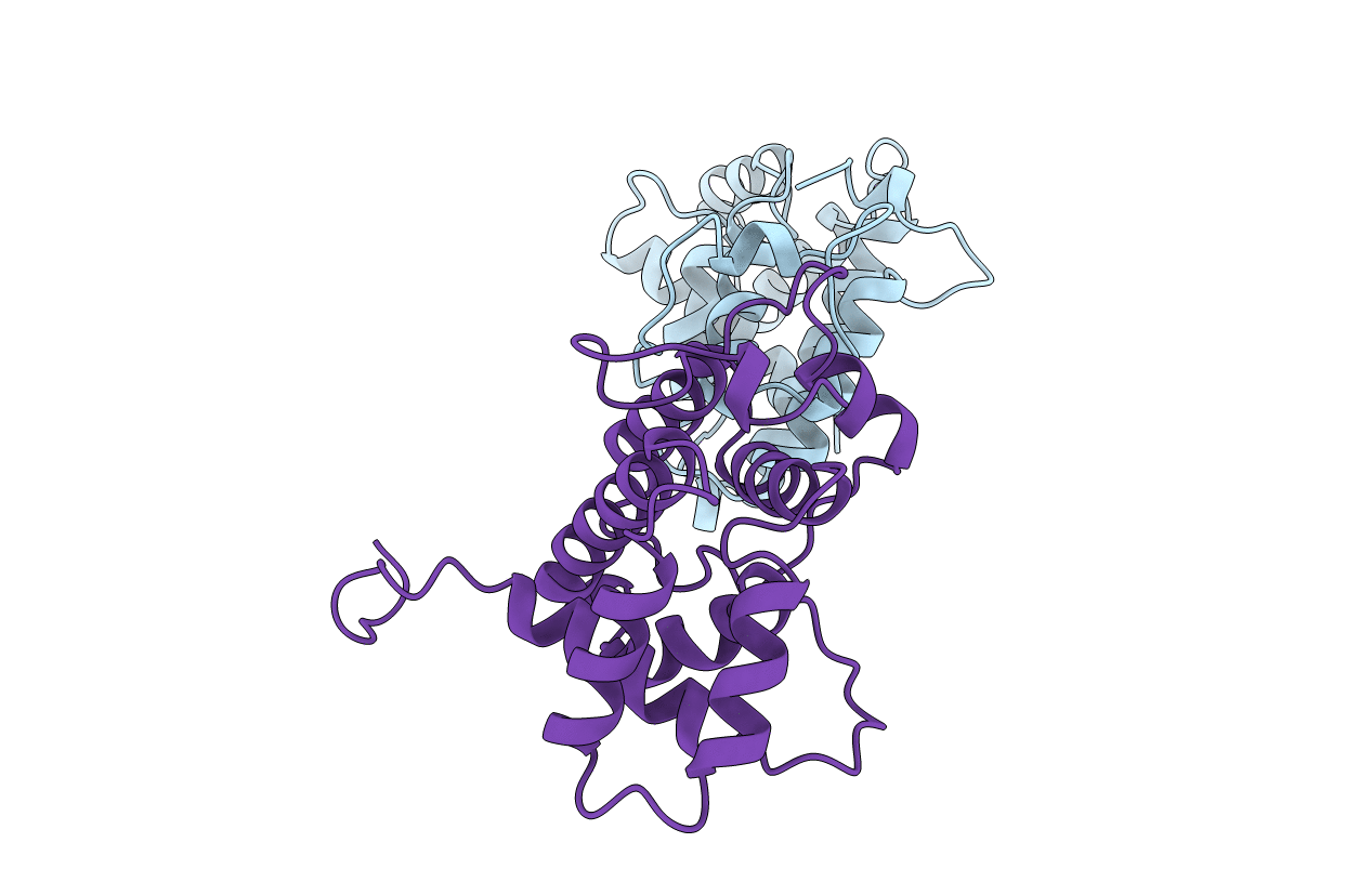
1PBW image
Deposition Date 1996-10-17
Release Date 1997-03-12
Last Version Date 2024-02-14
Entry Detail
PDB ID:
1PBW
Title:
STRUCTURE OF BCR-HOMOLOGY (BH) DOMAIN
Biological Source:
Source Organism:
Homo sapiens (Taxon ID: 9606)
Host Organism:
Method Details:
Experimental Method:
Resolution:
2.00 Å
R-Value Free:
0.23
R-Value Work:
0.18
R-Value Observed:
0.18
Space Group:
P 1 21 1
Macromolecular Entities
Structures with similar UniProt ID
Protein Blast
Polymer Type:polypeptide(L)
Molecule:PHOSPHATIDYLINOSITOL 3-KINASE
Gene (Uniprot):PIK3R1
Chain IDs:A, B
Chain Length:216
Number of Molecules:2
Biological Source:Homo sapiens
Primary Citation
Crystal structure of the breakpoint cluster region-homology domain from phosphoinositide 3-kinase p85 alpha subunit.
Proc.Natl.Acad.Sci.USA 93 14373 14378 (1996)
PMID: 8962058 DOI: 10.1073/pnas.93.25.14373

Abstact

Proteins such as the product of the break-point cluster region, chimaerin, and the Src homology 3-binding protein 3BP1, are GTPase activating proteins (GAPs) for members of the Rho subfamily of small GTP-binding proteins (G proteins or GTPases). A 200-residue region, named the breakpoint cluster region-homology (BH) domain, is responsible for the GAP activity. We describe here the crystal structure of the BH domain from the p85 subunit of phosphatidylinositol 3-kinase at 2.0 A resolution. The domain is composed of seven helices, having a previously unobserved arrangement. A core of four helices contains most residues that are conserved in the BH family. Their packing suggests the location of a G-protein binding site. This structure of a GAP-like domain for small GTP-binding proteins provides a framework for analyzing the function of this class of molecules.

Legend

Protein

Chemical

Disease

Primary Citation of related structures