1PA7 image
Entry Detail
PDB ID:
1PA7
Title:
Crystal structure of amino-terminal microtubule binding domain of EB1
Biological Source:
Source Organism:
PDB Version:
Deposition Date:
2003-05-13
Release Date:
2003-10-21
Method Details:
Experimental Method:
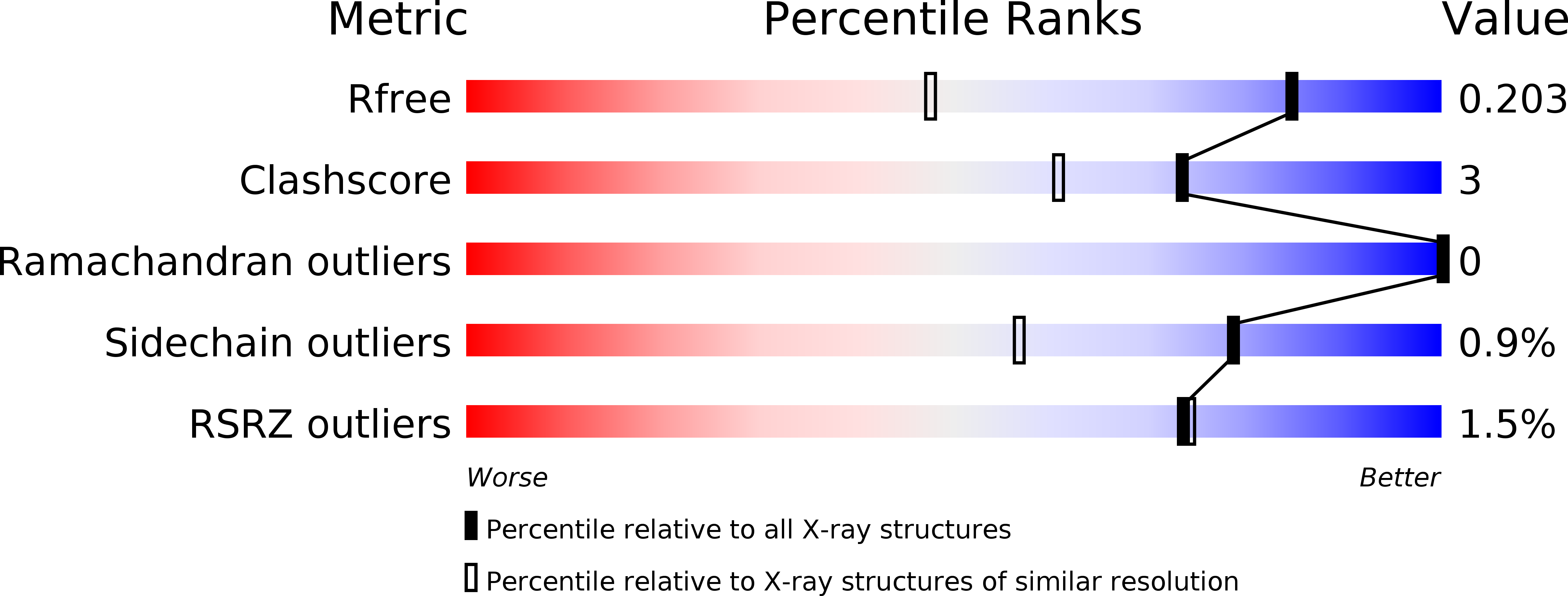
Resolution:
1.45 Å
R-Value Free:
0.18
R-Value Work:
0.17
R-Value Observed:
0.17
Space Group:
P 1 21 1
Macromolecular Entities
Polymer Type:polypeptide(L)
Description:Microtubule-associated protein RP/EB family member 1
Chain IDs:A
Chain Length:130
Number of Molecules:1
Biological Source:Homo sapiens
Ligand Molecules
Primary Citation
Crystal structure of the amino-terminal microtubule-binding domain of end-binding protein 1 (EB1)
J.Biol.Chem. 278 36430 36434 (2003)
PMID: 12857735 DOI: 10.1074/jbc.M305773200

Abstact

The end-binding protein 1 (EB1) family is a highly conserved group of proteins that localizes to the plus-ends of microtubules. EB1 has been shown to play an important role in regulating microtubule dynamics and chromosome segregation, but its regulation mechanism is poorly understood. We have determined the 1.45-A resolution crystal structure of the amino-terminal domain of EB1, which is essential for microtubule binding, and show that it forms a calponin homology (CH) domain fold that is found in many proteins involved in the actin cytoskeleton. The functional CH domain for actin binding is a tandem pair, whereas EB1 is the first example of a single CH domain that can associate with the microtubule filament. Although our biochemical study shows that microtubule binding of EB1 is electrostatic in part, our mutational analysis suggests that the hydrophobic network, which is partially exposed in our crystal structure, is also important for the association. We propose that, like other actin-binding CH domains, EB1 employs the hydrophobic interaction to bind to microtubules.

Legend

Protein

Chemical

Disease

Primary Citation of related structures