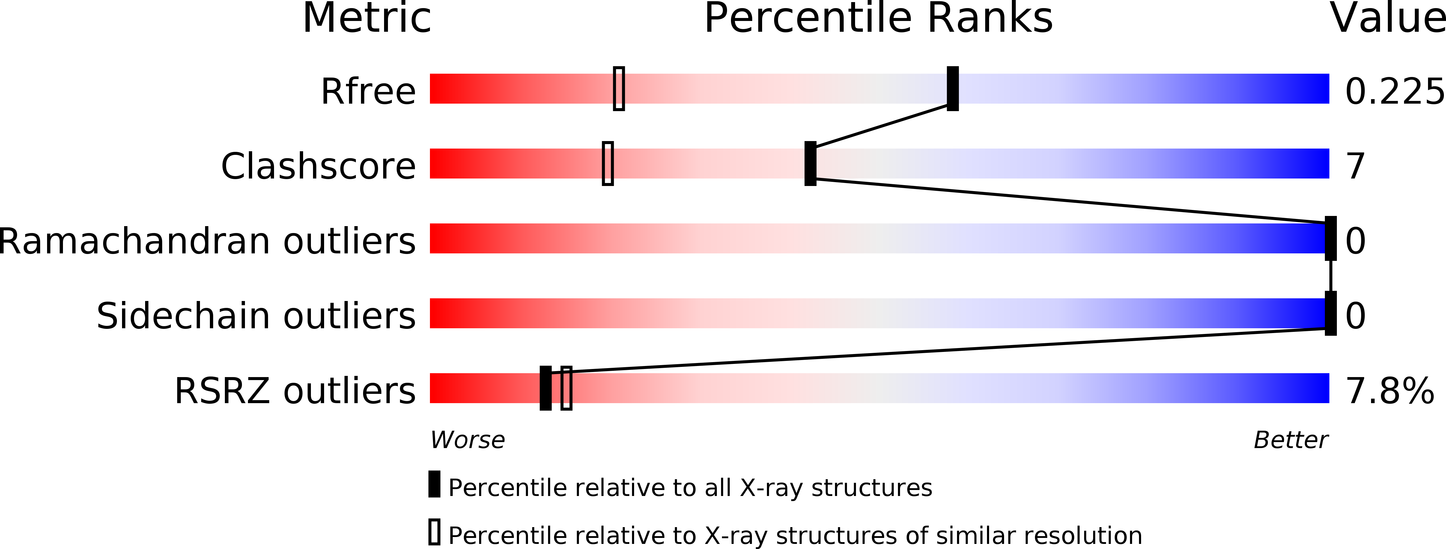
1P9H image
Deposition Date 2003-05-12
Release Date 2004-03-23
Last Version Date 2024-02-14
Entry Detail
PDB ID:
1P9H
Keywords:
Title:
CRYSTAL STRUCTURE OF THE COLLAGEN-BINDING DOMAIN OF YERSINIA ADHESIN YadA
Biological Source:
Source Organism:
Host Organism:
Method Details:
Experimental Method:
Resolution:
1.55 Å
R-Value Free:
0.20
R-Value Work:
0.19
Space Group:
H 3 2
Macromolecular Entities
Structures with similar UniProt ID
Protein Blast
Polymer Type:polypeptide(L)
Molecule:Invasin
Gene (Uniprot):yadA
Chain IDs:A
Chain Length:226
Number of Molecules:1
Biological Source:Yersinia enterocolitica
Primary Citation
The Yersinia adhesin YadA collagen-binding domain structure is a novel left-handed parallel beta-roll.
Embo J. 23 701 711 (2004)
PMID: 14765110 DOI: 10.1038/sj.emboj.7600100

Abstact

The crystal structure of the recombinant collagen-binding domain of Yersinia adhesin YadA from Yersinia enterocolitica serotype O:3 was solved at 1.55 A resolution. The trimeric structure is composed of head and neck regions, and the collagen binding head region is a novel nine-coiled left-handed parallel beta-roll. Before the beta-roll, the polypeptide loops from one monomer to the rest, and after the beta-roll the neck region does the same, making the transition from the globular head region to the narrower stalk domain. This creates an intrinsically stable 'lock nut' structure. The trimeric form of YadA is required for collagen binding, and mutagenesis of its surface residues allowed identification of a putative collagen-binding surface. Furthermore, a new structure-sequence motif for YadA beta-roll was used to identify putative YadA-head-like domains in a variety of human and plant pathogens. Such domains may therefore be a common bacterial strategy for avoiding host response.

Legend

Protein

Chemical

Disease

Primary Citation of related structures