1P99 image
Deposition Date 2003-05-09
Release Date 2004-01-20
Last Version Date 2024-02-14
Entry Detail
PDB ID:
1P99
Title:
1.7A crystal structure of protein PG110 from Staphylococcus aureus
Biological Source:
Method Details:
Experimental Method:
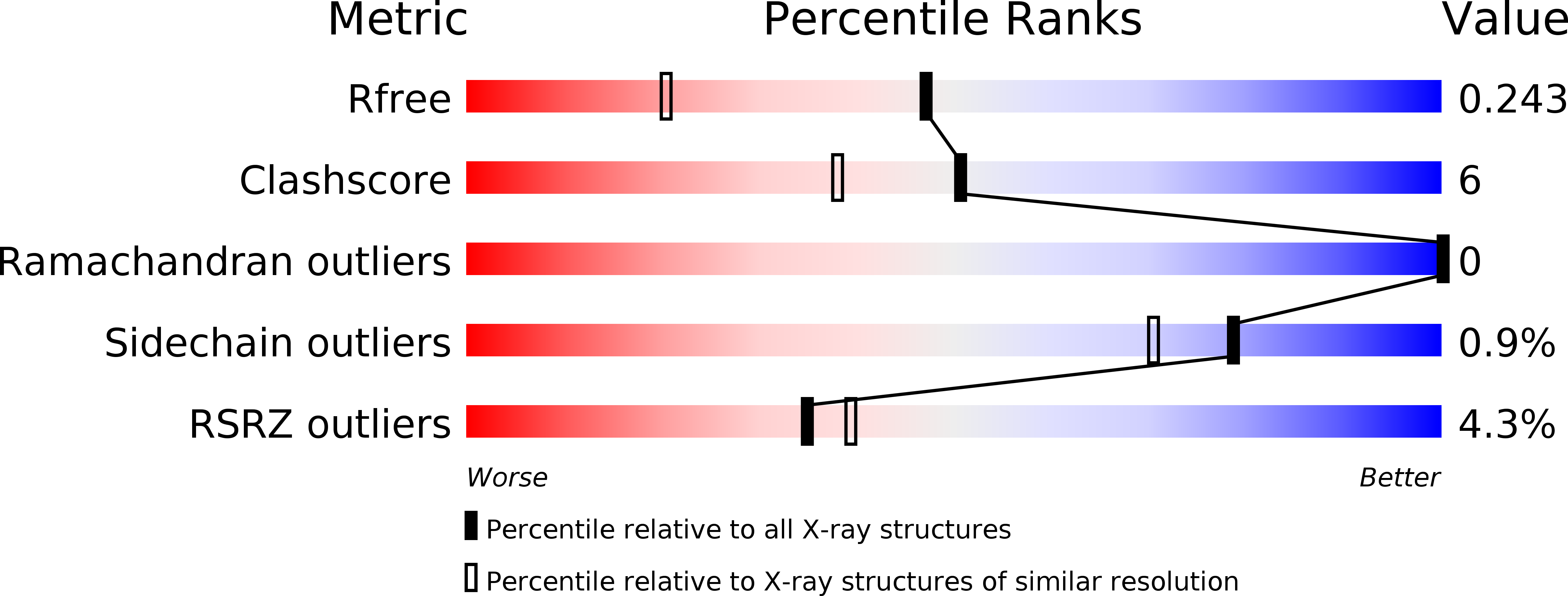
Resolution:
1.70 Å
R-Value Free:
0.23
R-Value Work:
0.2
R-Value Observed:
0.2
Space Group:
P 21 21 21
Macromolecular Entities
Structures with similar UniProt ID
Protein Blast
Polymer Type:polypeptide(L)
Molecule:Hypothetical protein PG110
Chain IDs:A
Chain Length:295
Number of Molecules:1
Biological Source:Staphylococcus aureus subsp. aureus Mu50
Primary Citation
The membrane-associated lipoprotein-9 GmpC from Staphylococcus aureus binds the dipeptide GlyMet via side chain interactions.
Biochemistry 43 16193 16202 (2004)
PMID: 15610013 DOI: 10.1021/bi048877o

Abstact

Bacterial dipeptide ABC transporters function to import a wide range of dipeptide substrates. This ability to transport a wide variety of dipeptides is conferred by the cognate substrate binding protein (SBP) of these transporters. SBPs bind dipeptides with little regard for their amino acid content. Here, we report the 1.7 A resolution structure of lipoprotein-9 (SA0422) of Staphylococcus aureus in complex with the dipeptide glycylmethionine. Experimental characterization of the subcellular location of the protein confirmed that SA0422 is an acylated, peripheral membrane protein. This is the first structure determined for an SBP of a Gram-positive dipeptide ABC transporter. Usually, binding of dipeptides occurs in a binding pocket that is largely hydrated and able to accommodate the side chains of several different amino acid residues. Unlike any other known SBP, lipoprotein-9 binds the side chains of the glycylmethionine dipeptide through very specific interactions. Lipoprotein-9 shares significant structural and sequence homology with the MetQ family of methionine SBP. Sequence comparisons between MetQ-like proteins and lipoprotein-9 suggest that the residues forming the tight interactions with the methionine side chains of the ligand are highly conserved between lipoprotein-9 and MetQ homologues, while the residues involved in coordinating the glycine residue are not. Modeling of the Vibrio cholerae MetQ and lipoprotein-9 binding pockets can account for lipoprotein-9 substrate specificity toward glycylmethionine. For this reason, we have designated lipoprotein-9 GmpC, for glycylmethionine binding protein.

Legend

Protein

Chemical

Disease

Primary Citation of related structures
Feedback Form
Name
Email
Institute
Feedback