
Deposition Date
2003-05-09
Release Date
2003-07-08
Last Version Date
2023-08-16
Entry Detail
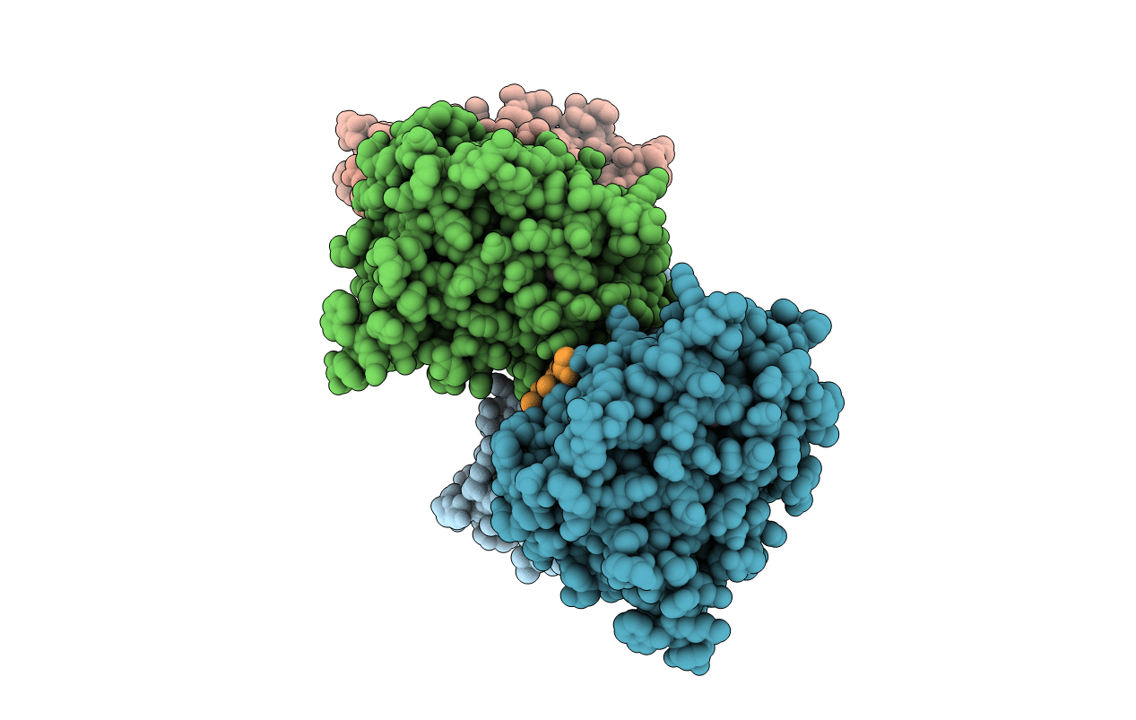
PDB ID:
1P93
Keywords:
Title:
CRYSTAL STRUCTURE OF THE AGONIST FORM OF GLUCOCORTICOID RECEPTOR
Biological Source:
Source Organism(s):
Homo sapiens (Taxon ID: 9606)
Expression System(s):
Method Details:
Experimental Method:
Resolution:
2.70 Å
R-Value Free:
0.36
R-Value Work:
0.34
R-Value Observed:
0.35
Space Group:
P 31
