1P8Z image
Deposition Date 2003-05-08
Release Date 2003-10-14
Last Version Date 2024-02-14
Entry Detail
PDB ID:
1P8Z
Title:
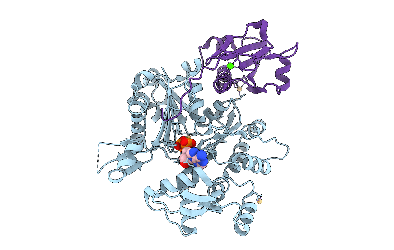
Complex Between Rabbit Muscle alpha-Actin: Human Gelsolin Residues Val26-Glu156
Biological Source:
Source Organism:
Host Organism:
Method Details:
Experimental Method:
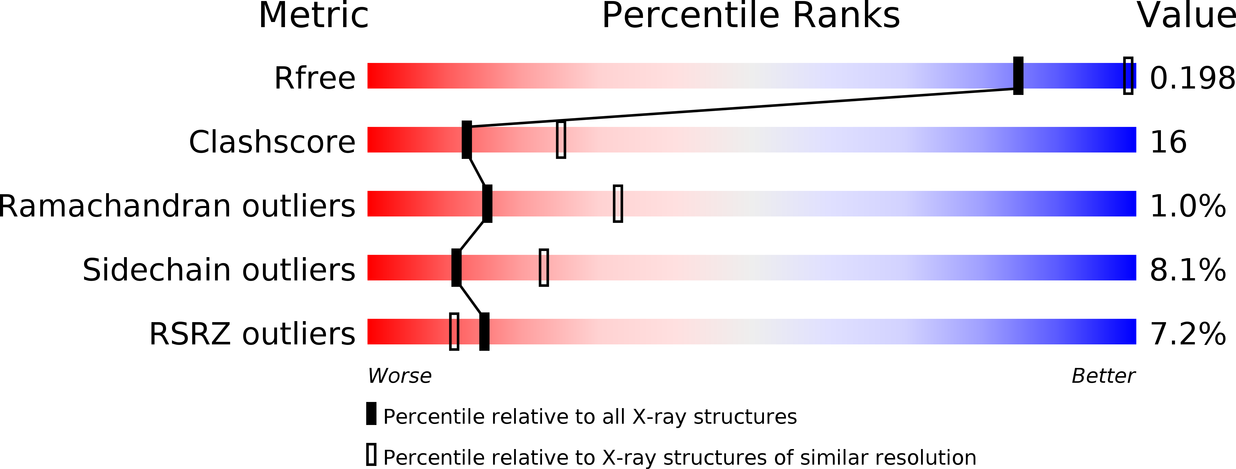
Resolution:
2.60 Å
R-Value Free:
0.26
R-Value Work:
0.20
R-Value Observed:
0.20
Space Group:
P 31 1 2
Macromolecular Entities
Structures with similar UniProt ID
Protein Blast
Polymer Type:polypeptide(L)
Molecule:Actin, alpha skeletal muscle
Gene (Uniprot):ACTA1
Chain IDs:B (auth: A)
Chain Length:377
Number of Molecules:1
Biological Source:Oryctolagus cuniculus
Structures with similar UniProt ID
Protein Blast
Polymer Type:polypeptide(L)
Molecule:Gelsolin precursor, plasma
Gene (Uniprot):GSN
Chain IDs:A (auth: G)
Chain Length:136
Number of Molecules:1
Biological Source:Homo sapiens
Primary Citation
From the First to the second domain of gelsolin: A common path on the surface of actin?
FEBS lett. 552 86 90 (2003)
PMID: 14527665 DOI: 10.1016/S0014-5793(03)00934-7

Abstact

We present the 2.6 A resolution crystal structure of a complex formed between G-actin and gelsolin fragment Met25-Gln160 (G1+). The structure differs from those of other gelsolin domain 1 (G1) complexes in that an additional six amino acid residues from the crucial linker region into gelsolin domain 2 (G2) are visible and are attached securely to the surface of actin. The linker segment extends away from G1 up the face of actin in a direction that infers G2 will bind along the same long-pitch helical strand as the actin bound to G1. This is consistent with a mechanism whereby G2 attaches gelsolin to the side of a filament and then directs G1 toward a position where it would disrupt actin-actin contacts. Alignment of the sequence of the structurally important residues within the G1-G2 linker with those of WH2 (WASp homology domain 2) domain protein family members (e.g. WASp (Wiscott-Aldridge syndrome protein) and thymosin beta4) suggests that the opposing activities of filament assembly and disassembly may exploit a common patch on the surface of actin.

Legend

Protein

Chemical

Disease

Primary Citation of related structures
Feedback Form
Name
Email
Institute
Feedback