1P8J image
Deposition Date 2003-05-07
Release Date 2003-07-08
Last Version Date 2025-03-26
Entry Detail
PDB ID:
1P8J
Title:
CRYSTAL STRUCTURE OF THE PROPROTEIN CONVERTASE FURIN
Biological Source:
Source Organism:
Mus musculus (Taxon ID: 10090)
(Taxon ID: )
Host Organism:
Method Details:
Experimental Method:
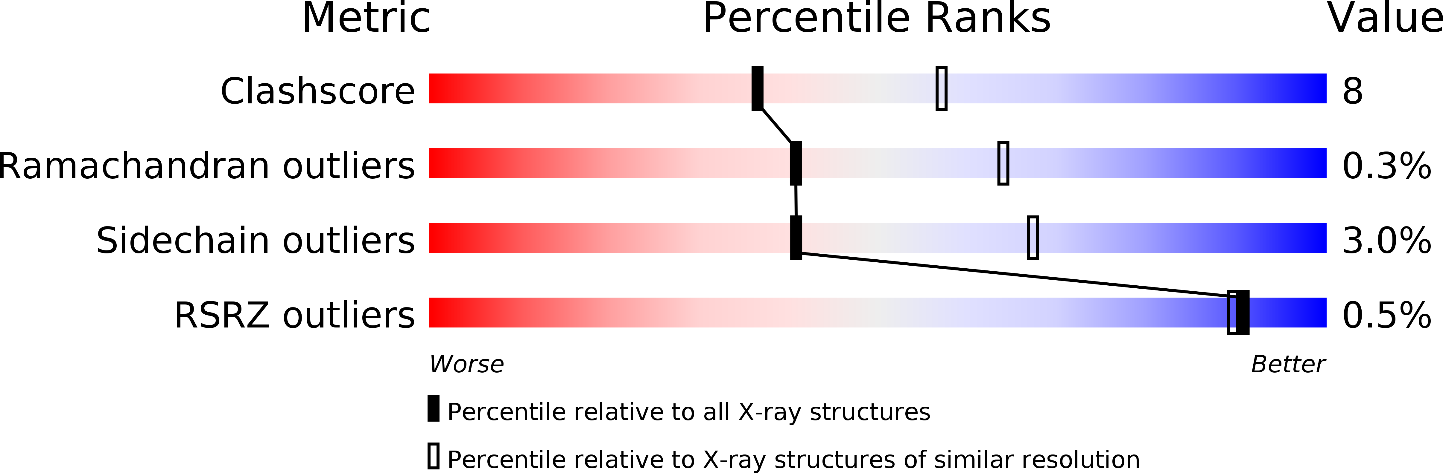
Resolution:
2.60 Å
R-Value Free:
0.21
R-Value Work:
0.18
R-Value Observed:
0.18
Space Group:
P 1
Macromolecular Entities
Polymer Type:polypeptide(L)
Molecule:Furin precursor
Gene (Uniprot):Furin
Chain IDs:A, B, C, D, E, F, G, H
Chain Length:471
Number of Molecules:8
Biological Source:Mus musculus
Polymer Type:polypeptide(L)
Molecule:DECANOYL-ARG-VAL-LYS-ARG-CHLOROMETHYLKETONE INHIBITOR
Chain IDs:I (auth: J), J (auth: K), K (auth: L), L (auth: M), M (auth: N), N (auth: P), O (auth: Q), P (auth: R)
Chain Length:6
Number of Molecules:8
Biological Source:
Modified Residue
Compound ID Chain ID Parent Comp ID Details 2D Image
AR7 I ARG ?
ASN A ASN GLYCOSYLATION SITE
Peptide-like Molecules
PRD_000278
Primary Citation
The Crystal Structure of the Proprotein Processing Proteinase Furin Explains its Stringent Specificity
Nat.Struct.Biol. 10 520 526 (2003)
PMID: 12794637 DOI: 10.1038/nsb941

Abstact

In eukaryotes, many essential secreted proteins and peptide hormones are excised from larger precursors by members of a class of calcium-dependent endoproteinases, the prohormone-proprotein convertases (PCs). Furin, the best-characterized member of the mammalian PC family, has essential functions in embryogenesis and homeostasis but is also implicated in various pathologies such as tumor metastasis, neurodegeneration and various bacterial and viral diseases caused by such pathogens as anthrax and pathogenic Ebola virus strains. Furin cleaves protein precursors with narrow specificity following basic Arg-Xaa-Lys/Arg-Arg-like motifs. The 2.6 A crystal structure of the decanoyl-Arg-Val-Lys-Arg-chloromethylketone (dec-RVKR-cmk)-inhibited mouse furin ectodomain, the first PC structure, reveals an eight-stranded jelly-roll P domain associated with the catalytic domain. Contoured surface loops shape the active site by cleft, thus explaining furin's stringent requirement for arginine at P1 and P4, and lysine at P2 sites by highly charge-complementary pockets. The structure also explains furin's preference for basic residues at P3, P5 and P6 sites. This structure will aid in the rational design of antiviral and antibacterial drugs.

Legend

Protein

Chemical

Disease

Primary Citation of related structures