1P8G image
Deposition Date 2003-05-07
Release Date 2003-11-25
Last Version Date 2024-05-22
Entry Detail
PDB ID:
1P8G
Keywords:
Title:
The solution structure of apo CopZ from Bacillus subtilis
Biological Source:
Source Organism:
Host Organism:
Method Details:
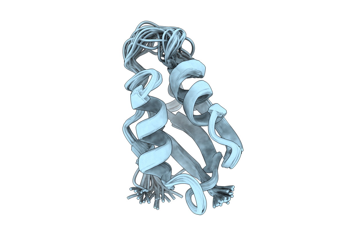
Experimental Method:
Conformers Calculated:
30
Conformers Submitted:
30
Selection Criteria:
structure with the lowest energy target function
Macromolecular Entities
Polymer Type:polypeptide(L)
Molecule:similar to mercuric transport protein
Gene (Uniprot):copZ
Chain IDs:A
Chain Length:73
Number of Molecules:1
Biological Source:Bacillus subtilis
Ligand Molecules
Primary Citation
Solution Structure of Apo CopZ from Bacillus subtilis: Further Analysis of the Changes Associated with the Presence of Copper
Biochemistry 42 13422 13428 (2003)
PMID: 14621987 DOI: 10.1021/bi0353326

Abstact

The solution structure of apo CopZ from Bacillus subtilis has been determined with the aim of investigating the changes in the hydrophobic interactions around the M-X-C-X-X-C copper(I) binding motif upon metal binding. The methionine of this motif (Met 11 in CopZ) points toward the solvent in apo CopZ, whereas its sulfur atom is close to the metal ion in the metal-loaded protein, though probably not at binding distance. This change is associated with the weakening of the interaction between Leu 37 and Cys 16, present in the apo form, and the formation of an interaction between Met 11 and Tyr 65. Loops 1, 3, and 5 are affected by metal binding. Comparison with the structure of other homologous proteins confirms that often metal binding affects a hydrophobic patch around the metal site, possibly for optimizing and tuning the hydrophobic interactions with the partners. It is also shown that copper(I) exchanges among apo CopZ molecules in slow exchange on the NMR time scale, whereas it is known that such exchange between partner molecules (i.e., metallochaperones and metal pumps) is fast.

Legend

Protein

Chemical

Disease

Primary Citation of related structures