1P7H image
Deposition Date 2003-05-01
Release Date 2003-09-23
Last Version Date 2024-02-14
Entry Detail
PDB ID:
1P7H
Title:
Structure of NFAT1 bound as a dimer to the HIV-1 LTR kB element
Biological Source:
Source Organism:
Homo sapiens (Taxon ID: 9606)
Method Details:
Experimental Method:
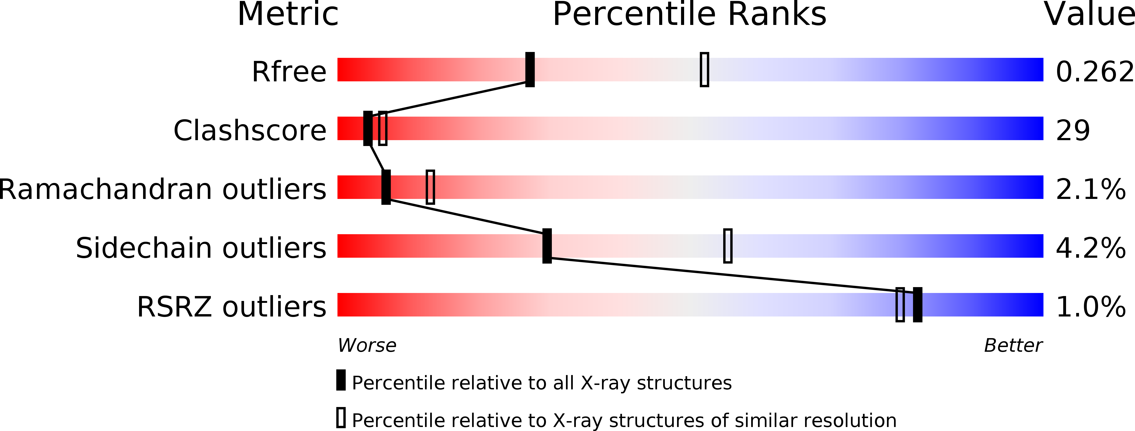
Resolution:
2.60 Å
R-Value Free:
0.26
R-Value Work:
0.23
R-Value Observed:
0.23
Space Group:
P 1
Macromolecular Entities
Structures with similar UniProt ID
Protein Blast
Polymer Type:polypeptide(L)
Molecule:Nuclear factor of activated T-cells, cytoplasmic 2
Gene (Uniprot):NFATC2
Chain IDs:E (auth: L), F (auth: M), G (auth: N), H (auth: O)
Chain Length:286
Number of Molecules:4
Biological Source:Homo sapiens
Primary Citation
Structure of NFAT1 bound as a dimer to the HIV-1 LTR kappa B element
Nat.Struct.Biol. 10 800 806 (2003)
PMID: 12949493 DOI: 10.1038/nsb981

Abstact

DNA binding by NFAT1 as a dimer has been implicated in the activation of host and viral genes. Here we report a crystal structure of NFAT1 bound cooperatively as a dimer to the highly conserved kappa B site from the human immunodeficiency virus 1 (HIV-1) long terminal repeat (LTR). This structure reveals a new mode of dimerization and protein-DNA recognition by the Rel homology region (RHR) of NFAT1. The two NFAT1 monomers form a complete circle around the kappa B DNA through protein-protein interactions mediated by both their N- and C-terminal subdomains. The major dimer interface, formed by the C-terminal domain, is asymmetric and substantially different from the symmetric dimer interface seen in other Rel family proteins. Comparison to other NFAT structures, including NFAT5 and the NFAT1-Fos-Jun-ARRE2 complex, reveals that NFAT1 adopts different conformations and its protein surfaces mediate distinct protein-protein interactions in the context of different DNA sites.

Legend

Protein

Chemical

Disease

Primary Citation of related structures