1P6O image
Deposition Date 2003-04-29
Release Date 2003-08-19
Last Version Date 2023-08-16
Entry Detail
PDB ID:
1P6O
Keywords:
Title:
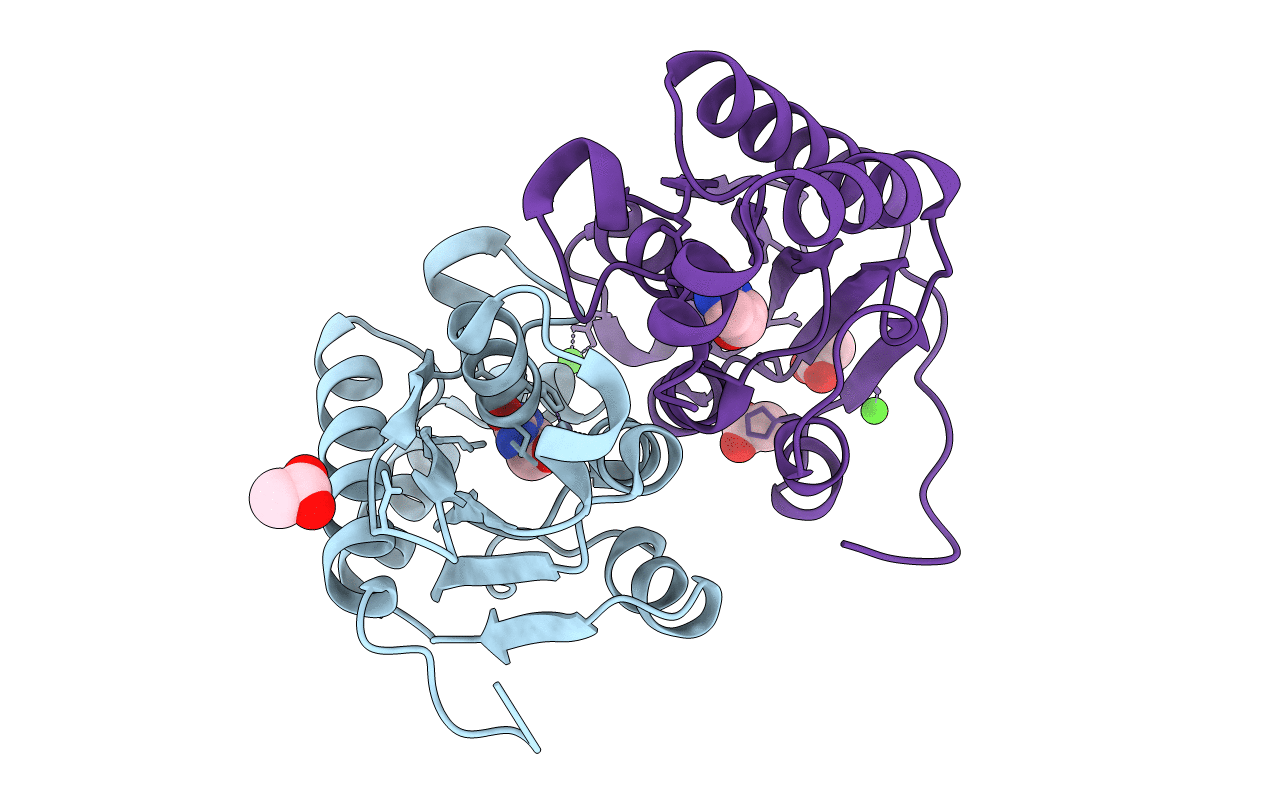
The crystal structure of yeast cytosine deaminase bound to 4(R)-hydroxyl-3,4-dihydropyrimidine at 1.14 angstroms.
Biological Source:
Source Organism:
Host Organism:
Method Details:
Experimental Method:
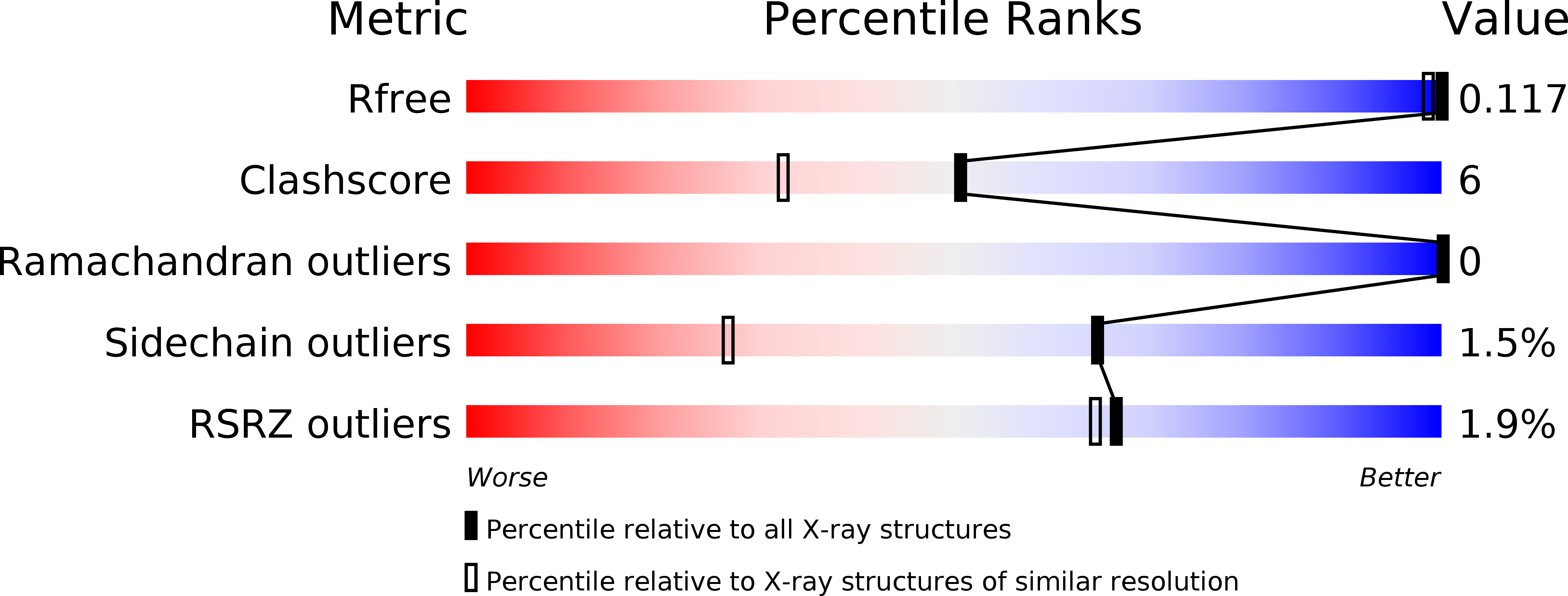
Resolution:
1.14 Å
R-Value Free:
0.15
R-Value Work:
0.10
R-Value Observed:
0.10
Space Group:
P 21 21 21
Macromolecular Entities
Structures with similar UniProt ID
Protein Blast
Polymer Type:polypeptide(L)
Molecule:Cytosine deaminase
Gene (Uniprot):FCY1
Chain IDs:A, B
Chain Length:161
Number of Molecules:2
Biological Source:Saccharomyces cerevisiae
Primary Citation
The 1.14 a crystal structure of yeast Cytosine deaminase. Evolution of nucleotide salvage enzymes and implications for genetic chemotherapy.
Structure 11 961 972 (2003)
PMID: 12906827 DOI: 10.1016/S0969-2126(03)00153-9

Abstact

Cytosine deaminase (CD) catalyzes the deamination of cytosine and is only present in prokaryotes and fungi, where it is a member of the pyrimidine salvage pathway. The enzyme is of interest both for antimicrobial drug design and gene therapy applications against tumors. The structure of Saccharomyces cerevisiae CD has been determined in the presence and absence of a mechanism-based inhibitor, at 1.14 and 1.43 A resolution, respectively. The enzyme forms an alpha/beta fold similar to bacterial cytidine deaminase, but with no similarity to the alpha/beta barrel fold used by bacterial cytosine deaminase or mammalian adenosine deaminase. The structures observed for bacterial, fungal, and mammalian nucleic acid deaminases represent an example of the parallel evolution of two unique protein folds to carry out the same reaction on a diverse array of substrates.

Legend

Protein

Chemical

Disease

Primary Citation of related structures