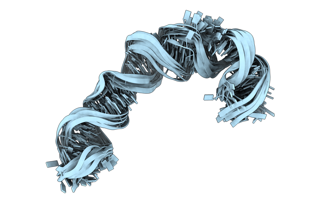
1P5O image
Deposition Date 2003-04-27
Release Date 2003-11-04
Last Version Date 2024-05-22
Entry Detail
PDB ID:
1P5O
Keywords:
Title:
Solution Structure of HCV IRES Domain II
Biological Source:
Source Organism:
(Taxon ID: )
Method Details:
Experimental Method:
Conformers Calculated:
200
Conformers Submitted:
12
Selection Criteria:
lowest restraint violation and total energy
Macromolecular Entities
Polymer Type:polyribonucleotide
Molecule:77-MER
Chain IDs:A
Chain Length:77
Number of Molecules:1
Biological Source:
Primary Citation
Structure of HCV IRES domain II determined by NMR.
Nat.Struct.Biol. 10 1033 1038 (2003)
PMID: 14578934 DOI: 10.1038/nsb1004

Abstact

Complex RNA structures regulate many biological processes, but are often too large for structure determination by NMR methods. The 5' untranslated region (5' UTR) of the hepatitis C viral (HCV) RNA genome contains an internal ribosome entry site (IRES) that binds to 40S ribosomal subunits with high affinity and specificity to control translation. Domain II of the HCV IRES forms a 25-kDa folded subdomain that may alter ribosome conformation. We report here the structure of domain II as determined using an NMR approach that combines short- and long-range structural data. Domain II adopts a distorted L-shape structure, and its overall shape in the free form is markedly similar to its 40S subunit-bound form; this suggests how domain II may modulate 40S subunit conformation. The results show how NMR can be used for structural analysis of large biological RNAs.

Legend

Protein

Chemical

Disease

Primary Citation of related structures