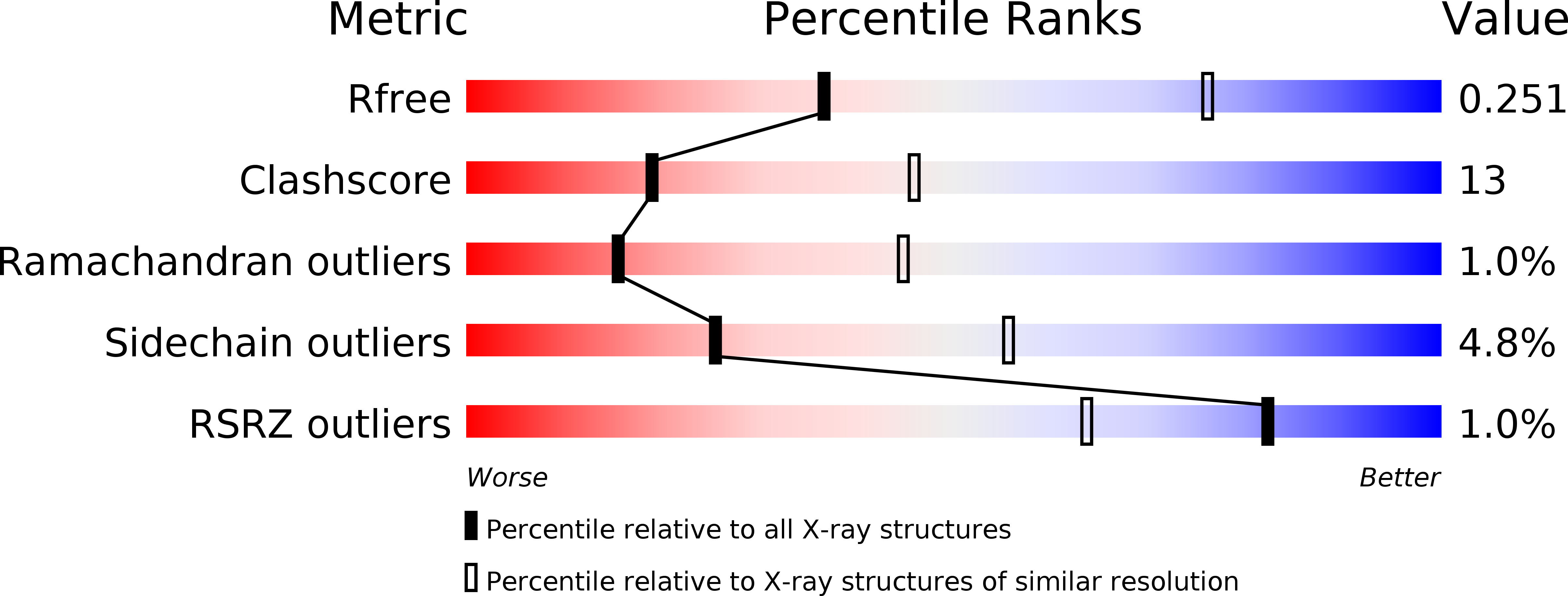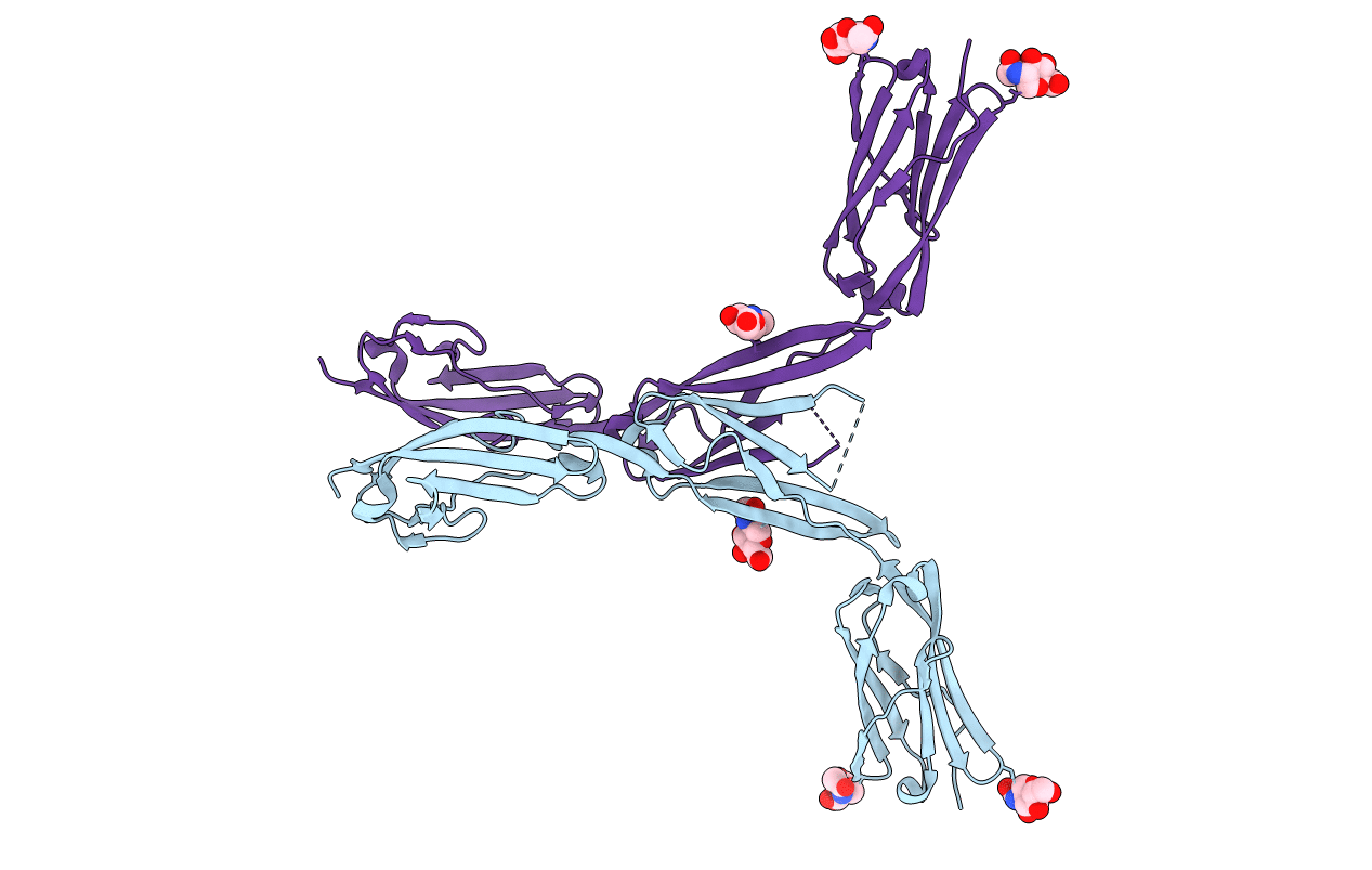
1P53 image
Deposition Date 2003-04-24
Release Date 2004-05-04
Last Version Date 2024-10-09
Entry Detail
PDB ID:
1P53
Keywords:
Title:
The Crystal Structure of ICAM-1 D3-D5 fragment
Biological Source:
Source Organism:
Homo sapiens (Taxon ID: 9606)
Host Organism:
Method Details:
Experimental Method:
Resolution:
3.06 Å
R-Value Free:
0.25
R-Value Work:
0.22
Space Group:
H 3 2
Macromolecular Entities
Structures with similar UniProt ID
Protein Blast
Polymer Type:polypeptide(L)
Molecule:Intercellular adhesion molecule-1
Gene (Uniprot):ICAM1
Chain IDs:A, B
Chain Length:266
Number of Molecules:2
Biological Source:Homo sapiens
Modified Residue
Compound ID Chain ID Parent Comp ID Details 2D Image
ASN A ASN GLYCOSYLATION SITE
Ligand Molecules
Primary Citation
Structural basis for dimerization of ICAM-1 on the cell surface.
Mol.Cell 14 269 276 (2004)
PMID: 15099525 DOI: 10.1016/S1097-2765(04)00204-7

Abstact

We have determined the 3.0 A crystal structure of the three C-terminal domains 3-5 (D3-D5) of ICAM-1. Combined with the previously known N-terminal two-domain structure (D1D2), a model of an entire ICAM-1 extracellular fragment has been constructed. This model should represent a general architecture of other ICAM family members, particularly ICAM-3 and ICAM-5. The observed intimate dimerization interaction at D4 and a stiff D4-D5 stem-like architecture provide a good structural explanation for the existence of preformed ICAM-1 cis dimers on the cell membrane. Together with another dimerization interface at D1, a band-like one-dimensional linear cluster of ICAM-1 on an antigen-presenting cell (APC) surface can be envisioned, which might explain the formation of an immunological synapse between an activated T cell and APC which is critical for T cell receptor signaling.

Legend

Protein

Chemical

Disease

Primary Citation of related structures