1P4X image
Deposition Date 2003-04-24
Release Date 2003-07-08
Last Version Date 2024-02-14
Entry Detail
PDB ID:
1P4X
Keywords:
Title:
Crystal structure of SarS protein from Staphylococcus Aureus
Biological Source:
Source Organism:
Host Organism:
Method Details:
Experimental Method:
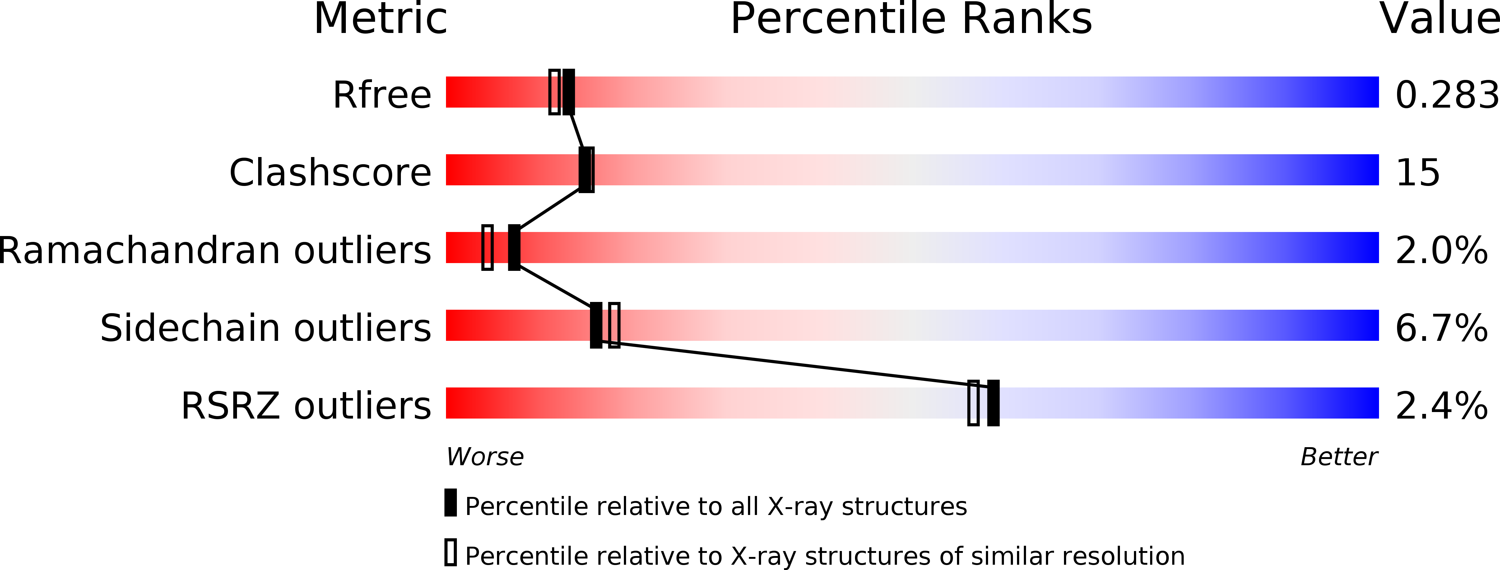
Resolution:
2.20 Å
R-Value Free:
0.28
R-Value Work:
0.24
R-Value Observed:
0.24
Space Group:
P 65 2 2
Macromolecular Entities
Structures with similar UniProt ID
Protein Blast
Polymer Type:polypeptide(L)
Molecule:staphylococcal accessory regulator A homologue
Chain IDs:A
Chain Length:250
Number of Molecules:1
Biological Source:Staphylococcus aureus
Primary Citation
Crystal structure of the SarS protein from Staphylococcus aureus
J.BACTERIOL. 185 4219 4225 (2003)
PMID: 12837797 DOI: 10.1128/JB.185.14.4219-4225.2003

Abstact

The expression of virulence determinants in Staphylococcus aureus is controlled by global regulatory loci (e.g., sarA and agr). One of these determinants, protein A (spa), is activated by sarS, which encodes a 250-residue DNA-binding protein. Genetic analysis indicated that the agr locus likely mediates spa repression by suppressing the transcription of sarS. Contrary to SarA and SarR, which require homodimer formation for proper function, SarS is unusual within the SarA protein family in that it contains two homologous halves, with each half sharing sequence similarity to SarA and SarR. Here we report the 2.2 A resolution X-ray crystal structure of the SarS protein. SarS has folds similar to those of SarR and, quite plausibly, the native SarA structure. Two typical winged-helix DNA-binding domains are connected by a well-ordered loop. The interactions between the two domains are extensive and conserved. The putative DNA-binding surface is highly positively charged. In contrast, negatively charged patches are located opposite to the DNA-binding surface. Furthermore, sequence alignment and structural comparison revealed that MarR has folds similar to those of SarR and SarS. Members of the MarR protein family have previously been implicated in the negative regulation of an efflux pump involved in multiple antibiotic resistance in many gram-negative species. We propose that MarR also belongs to the winged-helix protein family and has a similar mode of DNA binding as SarR and SarS and possibly the entire SarA protein family member. Based on the structural differences of SarR, SarS, and MarR, we further classified these winged-helix proteins to three subfamilies, SarA, SarS, and MarR. Finally, a possible transcription regulation mechanism is proposed.

Legend

Protein

Chemical

Disease

Primary Citation of related structures