
Deposition Date
2003-04-22
Release Date
2003-10-28
Last Version Date
2023-08-16
Entry Detail
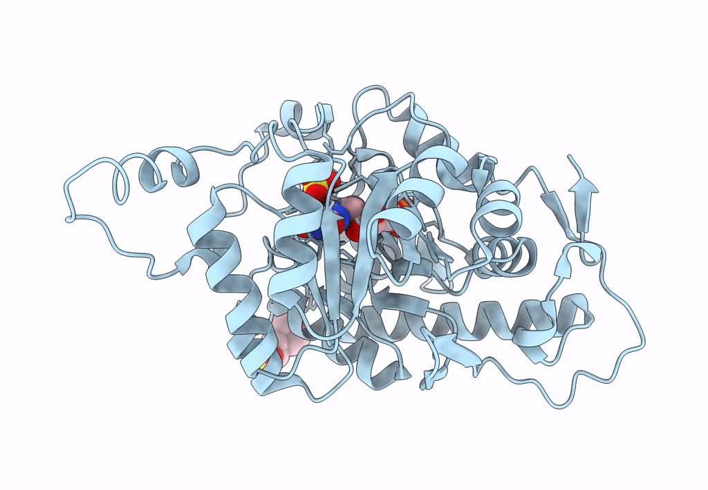
PDB ID:
1P4C
Keywords:
Title:
High Resolution Structure of Oxidized Active Mutant of (S)-Mandelate Dehydrogenase
Biological Source:
Source Organism(s):
Pseudomonas putida (Taxon ID: 303)
Spinacia oleracea (Taxon ID: 3562)
Spinacia oleracea (Taxon ID: 3562)
Expression System(s):
Method Details:
Experimental Method:
Resolution:
1.35 Å
R-Value Free:
0.19
R-Value Work:
0.18
R-Value Observed:
0.18
Space Group:
I 4
