
Deposition Date
2003-04-18
Release Date
2003-06-24
Last Version Date
2024-10-30
Entry Detail
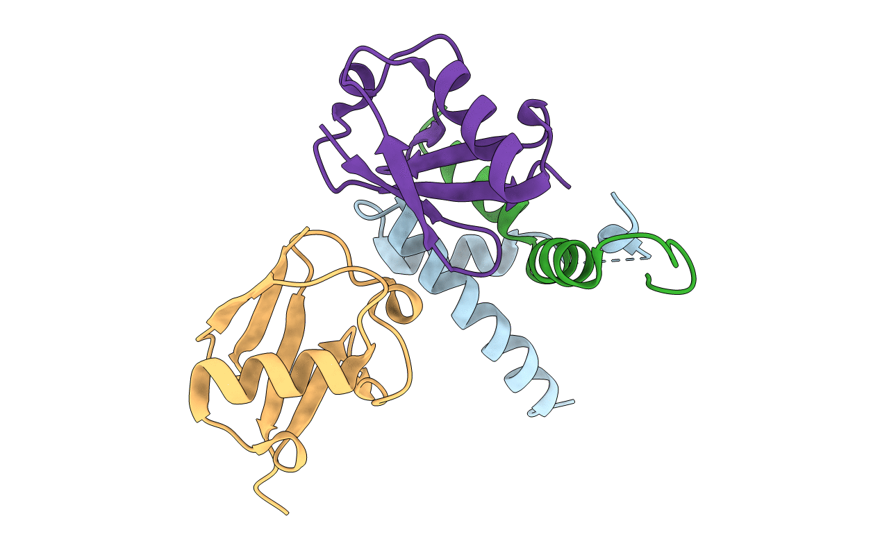
PDB ID:
1P3Q
Keywords:
Title:
Mechanism of Ubiquitin Recognition by the CUE Domain of VPS9
Biological Source:
Source Organism(s):
Saccharomyces cerevisiae (Taxon ID: 4932)
Bos taurus (Taxon ID: 9913)
Bos taurus (Taxon ID: 9913)
Expression System(s):
Method Details:
Experimental Method:
Resolution:
1.70 Å
R-Value Free:
0.27
R-Value Work:
0.26
Space Group:
C 1 2 1
