Planned Maintenance: Some services may turn out to be unavailable from 15th January, 2026 to 16th January, 2026. We apologize for the inconvenience!

1P0H image
Deposition Date 2003-04-10
Release Date 2003-09-09
Last Version Date 2024-02-14
Entry Detail
PDB ID:
1P0H
Keywords:
Title:
Crystal Structure of Rv0819 from Mycobacterium Tuberculosis MshD-Mycothiol Synthase Coenzyme A Complex
Biological Source:
Source Organism(s):
Expression System(s):
Method Details:
Experimental Method:
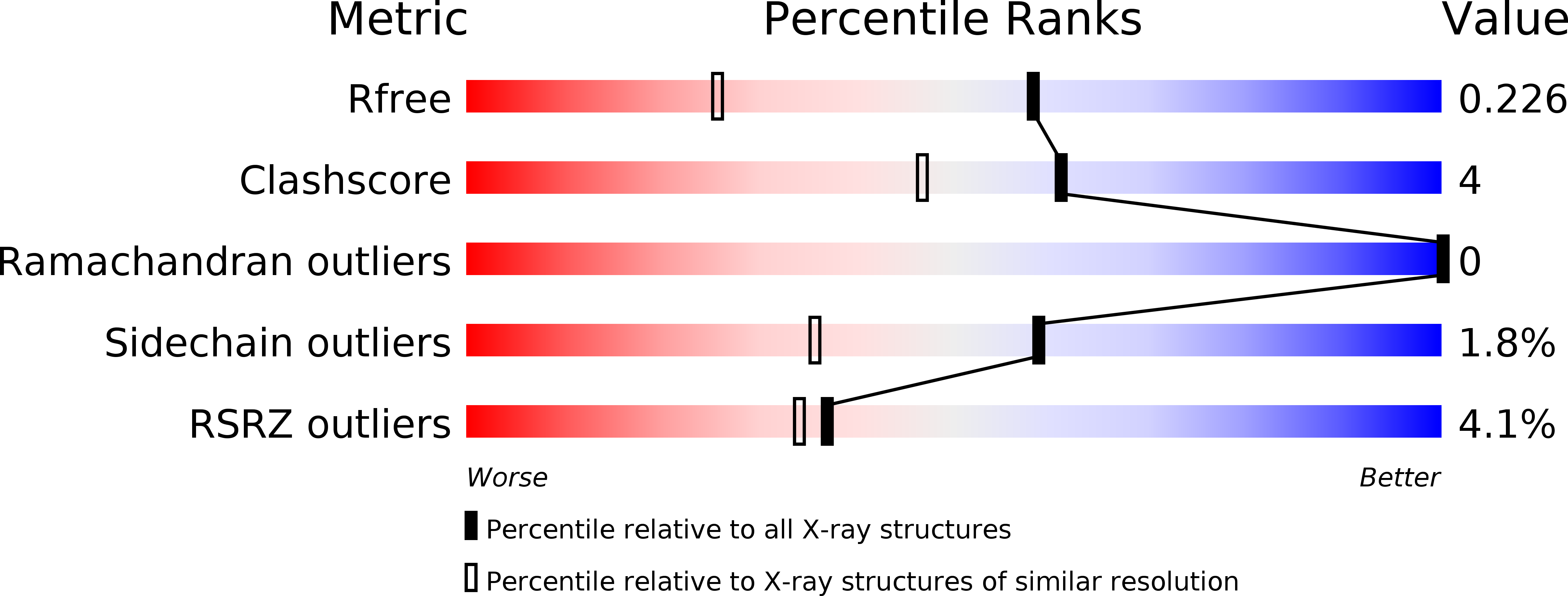
Resolution:
1.60 Å
R-Value Free:
0.23
R-Value Work:
0.19
R-Value Observed:
0.19
Space Group:
P 21 21 21
Macromolecular Entities
Structures with similar UniProt ID
Protein Blast
Polymer Type:polypeptide(L)
Molecule:hypothetical protein Rv0819
Chain IDs:A
Chain Length:318
Number of Molecules:1
Biological Source:Mycobacterium tuberculosis
Primary Citation
Crystal structure of mycothiol synthase (Rv0819) from Mycobacterium tuberculosis shows structural homology to the GNAT family of N-acetyltransferases.
Protein Sci. 12 1954 1959 (2003)
PMID: 12930994 DOI: 10.1110/ps.03153703

Abstact

Mycothiol is the predominant low-molecular weight thiol produced by actinomycetes, including Mycobacterium tuberculosis. The last reaction in the biosynthetic pathway for mycothiol is catalyzed by mycothiol synthase (MshD), which acetylates the cysteinyl amine of cysteine-glucosamine-inositol (Cys-GlcN-Ins). The crystal structure of MshD was determined in the presence of coenzyme A and acetyl-CoA. MshD consists of two tandem-repeated domains, each exhibiting the Gcn5-related N-acetyltransferase (GNAT) fold. These two domains superimpose with a root-mean-square deviation of 1.7 A over 88 residues, and each was found to bind one molecule of coenzyme, although the binding sites are quite different. The C-terminal domain has a similar active site to many GNAT members in which the acetyl group of the coenzyme is presented to an open active site slot. However, acetyl-CoA bound to the N-terminal domain is buried, and is apparently not positioned to promote acetyl transfer. A modeled substrate complex indicates that Cys-GlcN-Ins would only fill a portion of a negatively charged channel located between the two domains. This is the first structure determined for an enzyme involved in the biosynthesis of mycothiol.

Legend

Protein

Chemical

Disease

Primary Citation of related structures
Feedback Form
Name
Email
Institute
Feedback