1Oe0 image
Deposition Date 2003-03-17
Release Date 2003-10-23
Last Version Date 2023-12-13
Entry Detail
PDB ID:
1OE0
Keywords:
Title:
CRYSTAL STRUCTURE OF DROSOPHILA DEOXYRIBONUCLEOSIDE KINASE IN COMPLEX WITH DTTP
Biological Source:
Source Organism:
Host Organism:
Method Details:
Experimental Method:
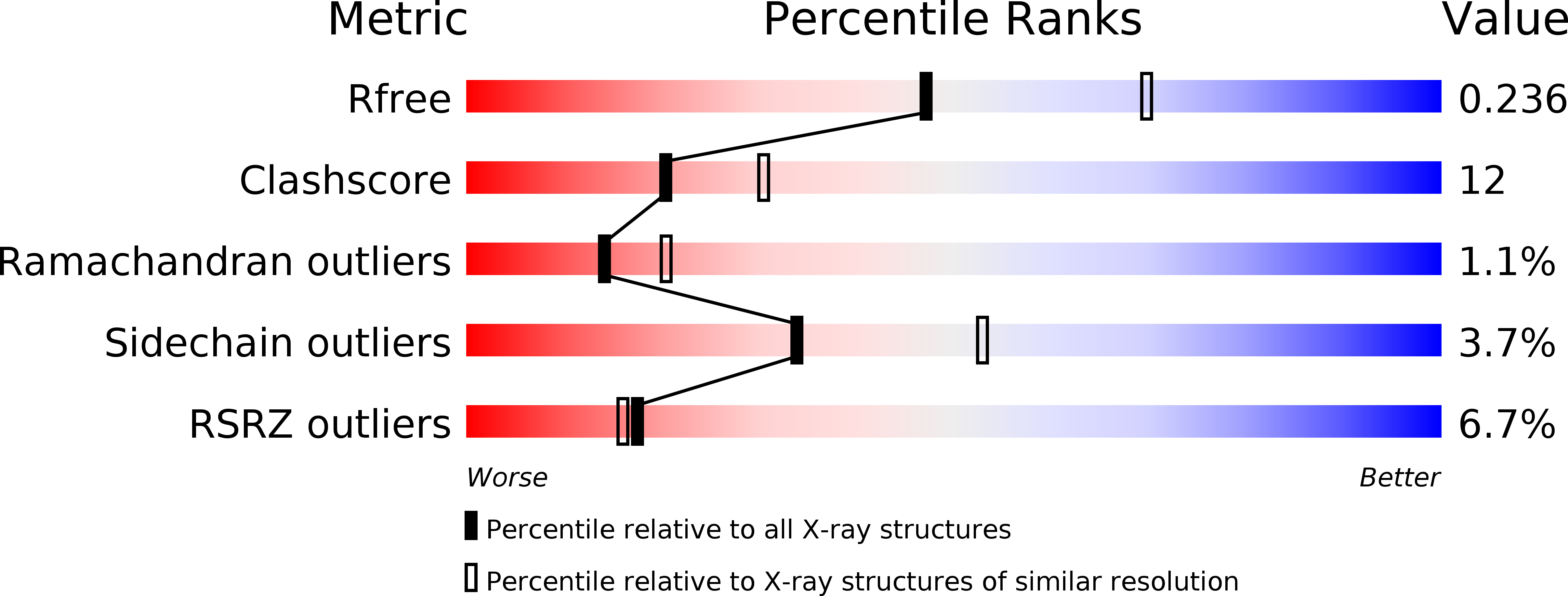
Resolution:
2.40 Å
R-Value Free:
0.24
R-Value Work:
0.22
R-Value Observed:
0.22
Space Group:
P 1 21 1
Macromolecular Entities
Polymer Type:polypeptide(L)
Molecule:DEOXYRIBONUCLEOSIDE KINASE
Gene (Uniprot):dnk
Chain IDs:A, B, C, D
Chain Length:230
Number of Molecules:4
Biological Source:DROSOPHILA MELANOGASTER
Primary Citation
Structural Basis for Feedback Inhibition of the Deoxyribonucleoside Salvage Pathway:Studies of the Drosophila Deoxyribonucleoside Kinase
Biochemistry 42 5706 ? (2003)
PMID: 12741827 DOI: 10.1021/BI0340043

Abstact

Deoxyribonucleoside kinases are feedback inhibited by the final products of the salvage pathway, the deoxyribonucleoside triphosphates. In the present study, the mechanism of feedback inhibition is presented based on the crystal structure of a complex between the fruit fly deoxyribonucleoside kinase and its feedback inhibitor deoxythymidine triphosphate. The inhibitor was found to be bound as a bisubstrate inhibitor with its nucleoside part in the nucleoside binding site and with its phosphate groups partially occupying the phosphate donor site. The overall structure of the enzyme--inhibitor complex is very similar to the enzyme--substrate complexes with deoxythymidine and deoxycytidine, except for a conformational change within a region otherwise directly involved in catalysis. This conformational change involves a magnesium ion, which is coordinated in the inhibitor complex to the phosphates and to the primary base, Glu52, that normally is positioned close to the 5'-OH of the substrate deoxyribose.

Legend

Protein

Chemical

Disease

Primary Citation of related structures