Deposition Date
2003-04-07
Release Date
2003-07-15
Last Version Date
2024-10-30
Entry Detail
PDB ID:
1OYV
Keywords:
Title:
Crystal structure of tomato inhibitor-II in a ternary complex with subtilisin Carlsberg
Biological Source:
Source Organism(s):
Bacillus licheniformis (Taxon ID: 1402)
Solanum lycopersicum (Taxon ID: 4081)
Solanum lycopersicum (Taxon ID: 4081)
Method Details:
Experimental Method:
Resolution:
2.50 Å
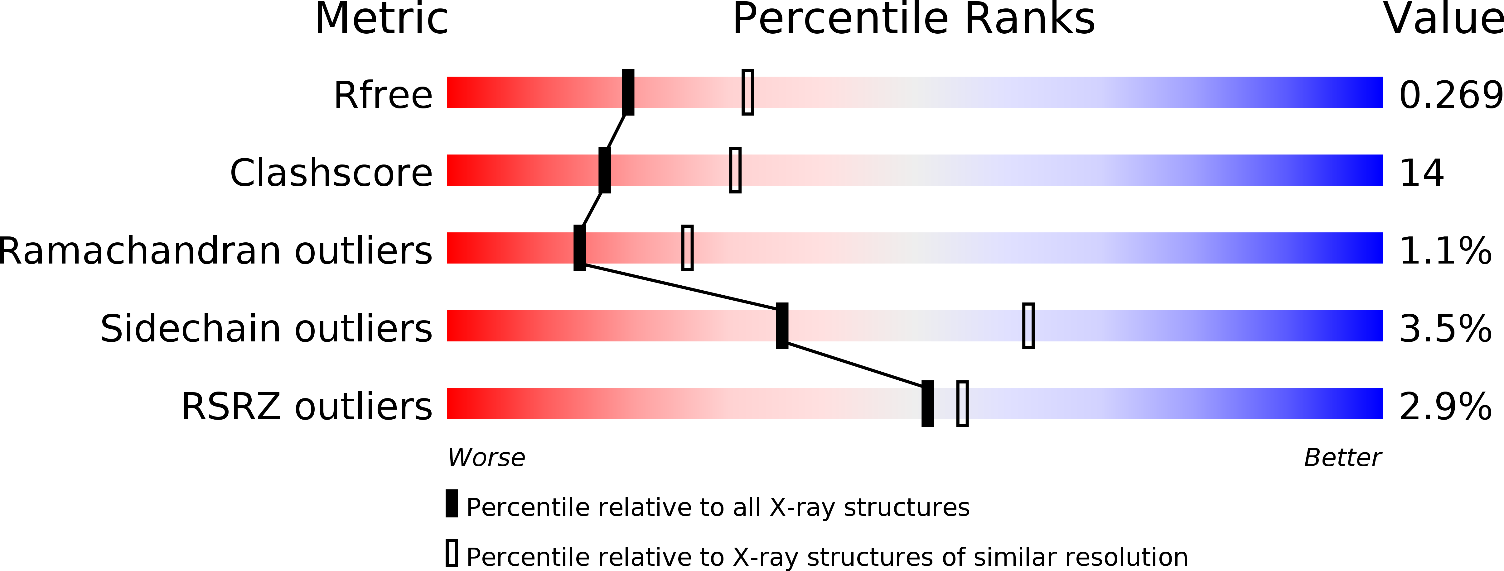
R-Value Free:
0.27
R-Value Work:
0.21
R-Value Observed:
0.22
Space Group:
C 1 2 1