1OYR image
Deposition Date 2003-04-07
Release Date 2004-03-09
Last Version Date 2024-02-14
Entry Detail
PDB ID:
1OYR
Keywords:
Title:
Crystal structure of the phosphorolytic exoribonuclease RNase PH from Bacillus subtilis
Biological Source:
Source Organism:
Method Details:
Experimental Method:
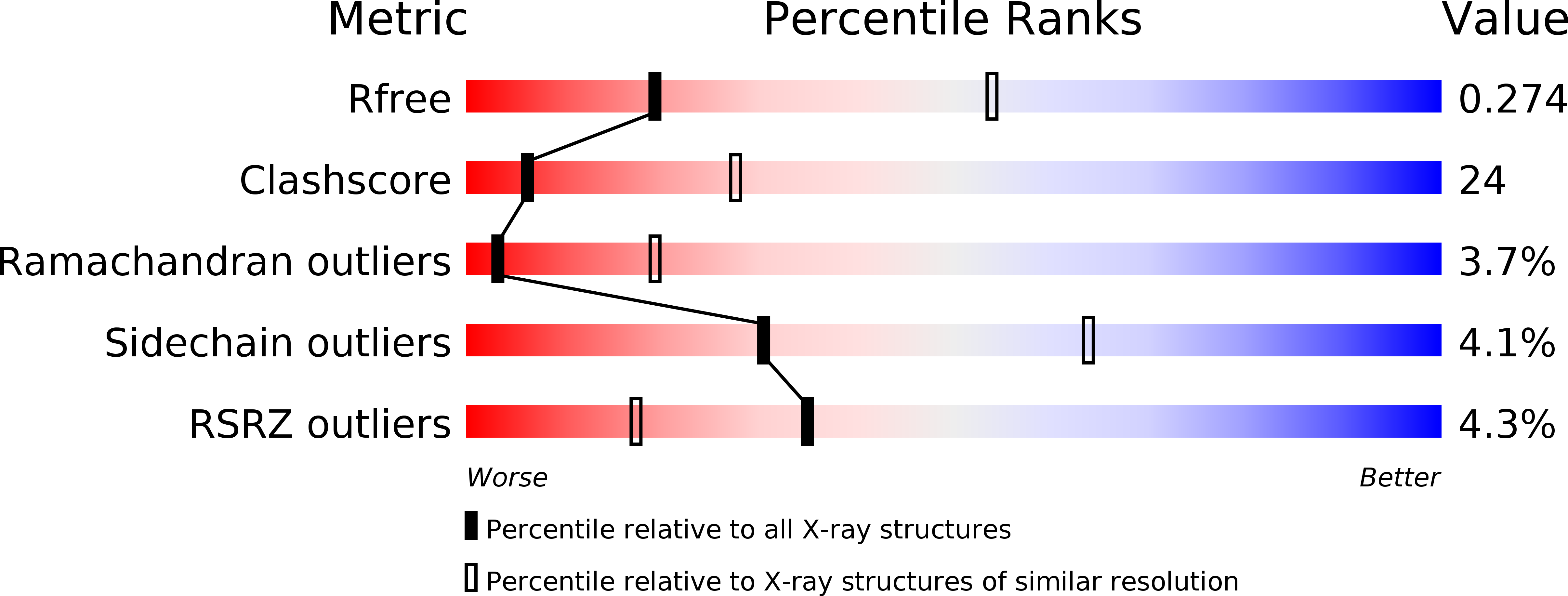
Resolution:
3.10 Å
R-Value Free:
0.28
R-Value Work:
0.27
Space Group:
P 21 21 21
Macromolecular Entities
Polymer Type:polypeptide(L)
Molecule:Ribonuclease PH
Gene (Uniprot):rph
Chain IDs:A, B, C, D, E, F
Chain Length:245
Number of Molecules:6
Biological Source:Bacillus subtilis
Primary Citation
Crystal structure of the phosphorolytic exoribonuclease RNase PH from Bacillus subtilis and implications for its quaternary structure and tRNA binding.
Protein Sci. 13 668 677 (2004)
PMID: 14767080 DOI: 10.1110/ps.03477004

Abstact

RNase PH is a member of the family of phosphorolytic 3' --> 5' exoribonucleases that also includes polynucleotide phosphorylase (PNPase). RNase PH is involved in the maturation of tRNA precursors and especially important for removal of nucleotide residues near the CCA acceptor end of the mature tRNAs. Wild-type and triple mutant R68Q-R73Q-R76Q RNase PH from Bacillus subtilis have been crystallized and the structures determined by X-ray diffraction to medium resolution. Wild-type and triple mutant RNase PH crystallize as a hexamer and dimer, respectively. The structures contain a rare left-handed beta alpha beta-motif in the N-terminal portion of the protein. This motif has also been identified in other enzymes involved in RNA metabolism. The RNase PH structure and active site can, despite low sequence similarity, be overlayed with the N-terminal core of the structure and active site of Streptomyces antibioticus PNPase. The surface of the RNase PH dimer fit the shape of a tRNA molecule.

Legend

Protein

Chemical

Disease

Primary Citation of related structures