1OYH image
Deposition Date 2003-04-04
Release Date 2004-04-13
Last Version Date 2024-10-30
Entry Detail
PDB ID:
1OYH
Keywords:
Title:
Crystal Structure of P13 Alanine Variant of Antithrombin
Biological Source:
Source Organism:
Homo sapiens (Taxon ID: 9606)
Host Organism:
Method Details:
Experimental Method:
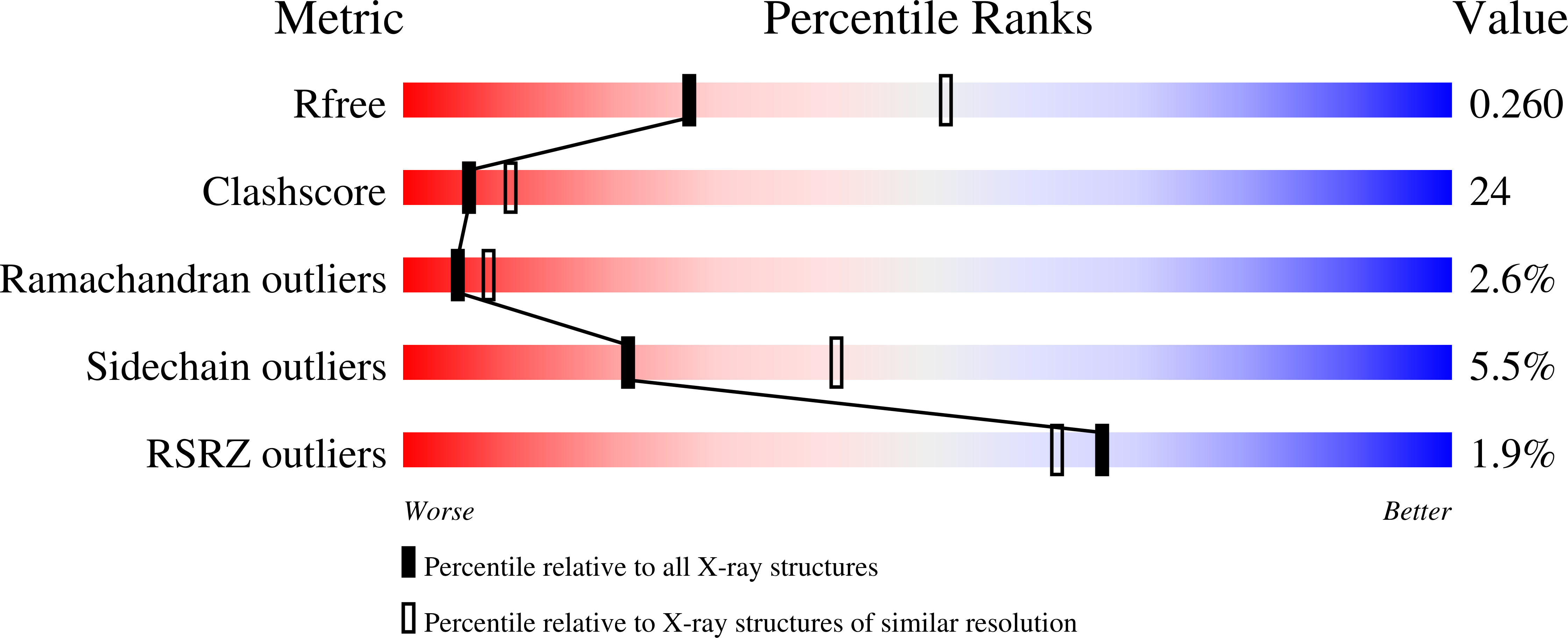
Resolution:
2.62 Å
R-Value Free:
0.25
R-Value Work:
0.22
Space Group:
P 1 21 1
Macromolecular Entities
Polymer Type:polypeptide(L)
Molecule:Antithrombin-III
Gene (Uniprot):SERPINC1
Mutations:E381A, S137A
Chain IDs:A (auth: I)
Chain Length:432
Number of Molecules:1
Biological Source:Homo sapiens
Polymer Type:polypeptide(L)
Molecule:Antithrombin-III
Gene (Uniprot):SERPINC1
Chain IDs:B (auth: L)
Chain Length:432
Number of Molecules:1
Biological Source:Homo sapiens
Modified Residue
Compound ID Chain ID Parent Comp ID Details 2D Image
ASN A ASN GLYCOSYLATION SITE
Ligand Molecules
Primary Citation
The influence of hinge region residue Glu-381 on antithrombin allostery and metastability
J.Biol.Chem. 279 4913 4921 (2004)
PMID: 14623882 DOI: 10.1074/jbc.M311644200

Abstact

Antithrombin becomes an efficient inhibitor of factor Xa and thrombin by binding a specific pentasaccharide sequence found on a small fraction of the heparan sulfate proteoglycans lining the microvaculature. In the structure of native antithrombin, the reactive center loop is restrained due to the insertion of its hinge region into the main beta-sheet A, whereas in the heparin-activated state the reactive center loop is freed from beta-sheet A. In both structures, hinge region residue Glu-381 makes several stabilizing contacts. To determine the role of these contacts in the allosteric mechanism of antithrombin activation, we replaced Glu-381 with an alanine. This variant is less active toward its target proteases than control antithrombin, due to a perturbation of the equilibrium between the two forms, and to an increase in stoichiometry of inhibition. Pentasaccharide binding affinity is reduced 4-fold due to an increase in the off-rate. These data suggest that the main role of Glu-381 is to stabilize the activated conformation. Stability studies also showed that the E381A variant is resistant to continued insertion of its reactive center loop upon incubation at 50 degrees C, suggesting new stabilizing interactions in the native structure. To test this hypothesis, and to aid in the interpretation of the kinetic data we solved to 2.6 A the structure of the variant. We conclude that wild-type Glu-381 interactions stabilize the activated state and decreases the energy barrier to full loop insertion.

Legend

Protein

Chemical

Disease

Primary Citation of related structures