1OX5 image
Entry Detail
PDB ID:
1OX5
Title:
TOWARDS UNDERSTANDING THE MECHANISM OF THE COMPLEX CYCLIZATION REACTION CATALYZED BY IMIDAZOLE GLYCEROPHOSPHATE SYNTHASE
Biological Source:
Source Organism:
Host Organism:
PDB Version:
Deposition Date:
2003-04-01
Release Date:
2003-06-17
Method Details:
Experimental Method:
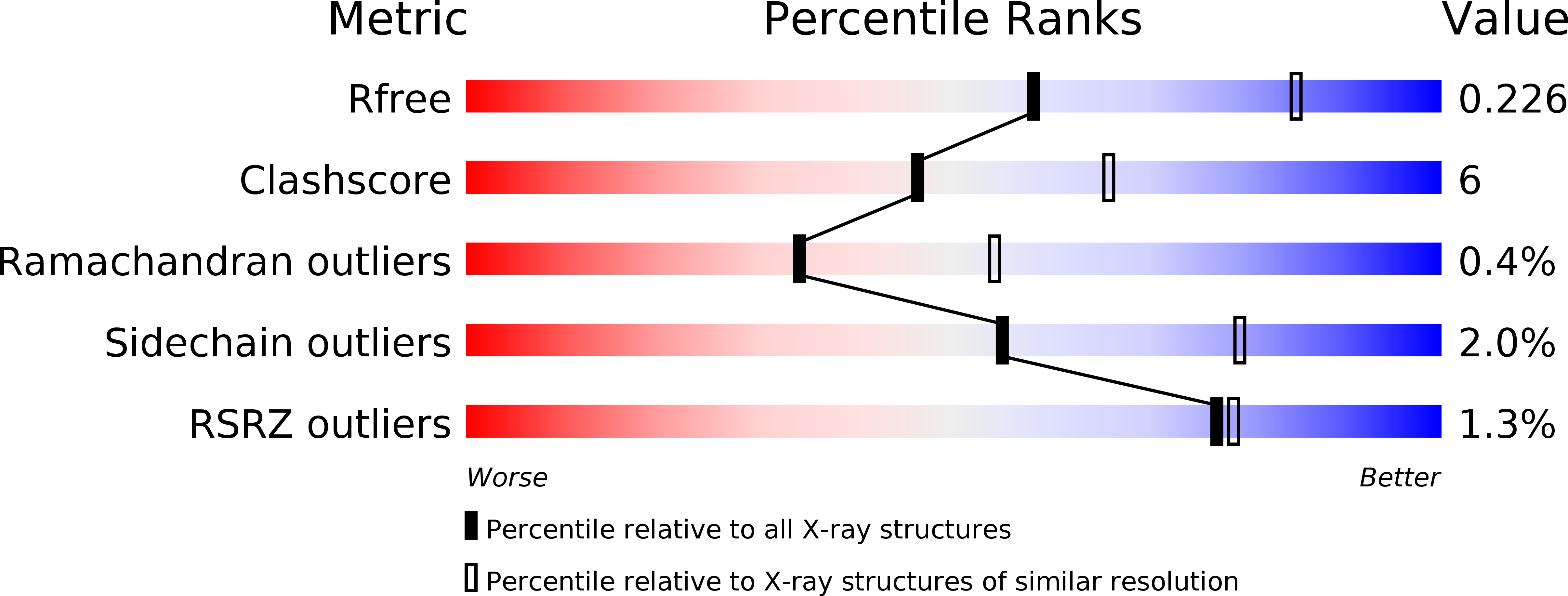
Resolution:
2.50 Å
R-Value Free:
0.24
R-Value Work:
0.22
Space Group:
P 21 21 21
Macromolecular Entities
Polymer Type:polypeptide(L)
Description:Imidazole glycerol phosphate synthase hisHF
Mutations:Cys83 residue is covalently modified by acivicin
Chain IDs:A, B
Chain Length:555
Number of Molecules:2
Biological Source:Saccharomyces cerevisiae
Modified Residue
Compound ID Chain ID Parent Comp ID Details 2D Image
5CS A CYS ?
Primary Citation
Towards Understanding the Mechanism of the Complex Cyclization Reaction Catalyzed by Imidazole Glycerophosphate Synthase:Crystal Structures of a Ternary Complex and the Free Enzyme
Biochemistry 42 7003 7012 (2003)
PMID: 12795595 DOI: 10.1021/bi034320h

Abstact

Imidazole glycerol phosphate synthase catalyzes formation of the imidazole ring in histidine biosynthesis. The enzyme is also a glutamine amidotransferase, which produces ammonia in a glutaminase active site and channels it through a 30-A internal tunnel to a cyclase active site. Glutaminase activity is impaired in the resting enzyme, and stimulated by substrate binding in the cyclase active site. The signaling mechanism was investigated in the crystal structure of a ternary complex in which the glutaminase active site was inactivated by a glutamine analogue and the unstable cyclase substrate was cryo-trapped in the active site. The orientation of N(1)-(5'-phosphoribulosyl)-formimino-5-aminoimidazole-4-carboxamide ribonucleotide in the cyclase active site implicates one side of the cyclase domain in signaling to the glutaminase domain. This side of the cyclase domain contains the interdomain hinge. Two interdomain hydrogen bonds, which do not exist in more open forms of the enzyme, are proposed as molecular signals. One hydrogen bond connects the cyclase domain to the substrate analogue in the glutaminase active site. The second hydrogen bond connects to a peptide that forms an oxyanion hole for stabilization of transient negative charge during glutamine hydrolysis. Peptide rearrangement induced by a fully closed domain interface is proposed to activate the glutaminase by unblocking the oxyanion hole. This interpretation is consistent with biochemical results [Myers, R. S., et al., (2003) Biochemistry 42, 7013-7022, the accompanying paper in this issue] and with structures of the free enzyme and a binary complex with a second glutamine analogue.

Legend

Protein

Chemical

Disease

Primary Citation of related structures