1OX3 image
Deposition Date 2003-04-01
Release Date 2004-04-13
Last Version Date 2023-08-16
Entry Detail
PDB ID:
1OX3
Keywords:
Title:
crystal structure of mini-fibritin
Biological Source:
Source Organism:
Method Details:
Experimental Method:
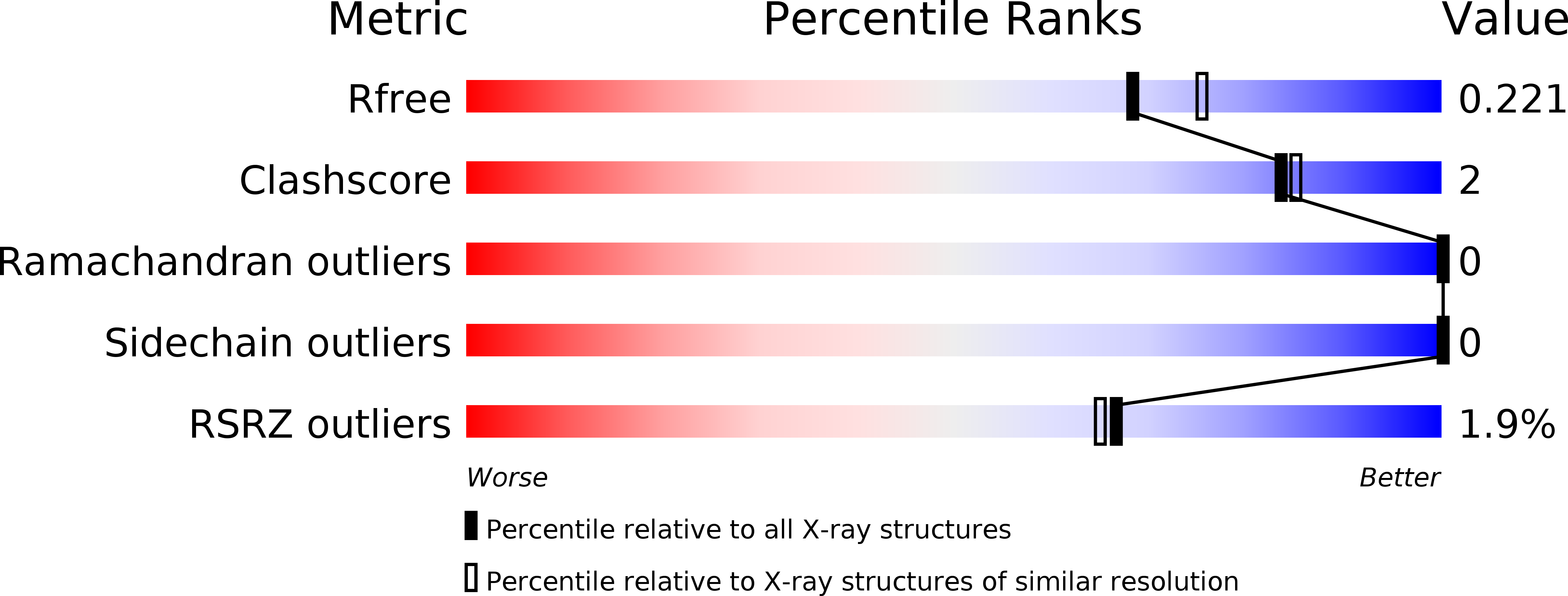
Resolution:
2.00 Å
R-Value Free:
0.22
R-Value Work:
0.20
R-Value Observed:
0.20
Space Group:
P 63
Macromolecular Entities
Polymer Type:polypeptide(L)
Molecule:Fibritin
Gene (Uniprot):wac
Chain IDs:A
Chain Length:109
Number of Molecules:1
Biological Source:Enterobacteria phage T4
Primary Citation
Design and Crystal Structure of Bacteriophage T4 Mini-Fibritin NCCF.
J.Mol.Biol. 339 927 935 (2004)
PMID: 15165860 DOI: 10.1016/j.jmb.2004.04.001

Abstact

Fibritin is a fibrous protein that forms "whiskers" attached to the neck of bacteriophage T4. Whiskers interact with the long tail fibers regulating the assembly and infectivity of the virus. The fibritin trimer includes the N-terminal domain responsible for attachment to the phage particle and for the collar formation, the central domain forming a 500 A long segmented coiled-coil structure, and the C-terminal "foldon" domain. We have designed a "mini" fibritin with most of the coiled-coil domain deleted, and solved its crystal structure. The non-helical N-terminal part represents a new protein fold that tightly interacts with the coiled-coil segment forming a single domain, as revealed by calorimetry. The analysis of the crystal structure and earlier electron microscopy data on the collar-whisker complex suggests the necessity of other proteins to participate in the collar formation. Crystal structure determination of the N-terminal domain of fibritin is the first step towards elucidating the detailed structure and assembly mechanism of the collar-whisker complex.

Legend

Protein

Chemical

Disease

Primary Citation of related structures