Deposition Date
2003-03-21
Release Date
2003-05-27
Last Version Date
2023-10-25
Entry Detail
PDB ID:
1OT3
Keywords:
Title:
Crystal structure of Drosophila deoxyribonucleotide kinase complexed with the substrate deoxythymidine
Biological Source:
Source Organism(s):
Drosophila melanogaster (Taxon ID: 7227)
Expression System(s):
Method Details:
Experimental Method:
Resolution:
2.50 Å
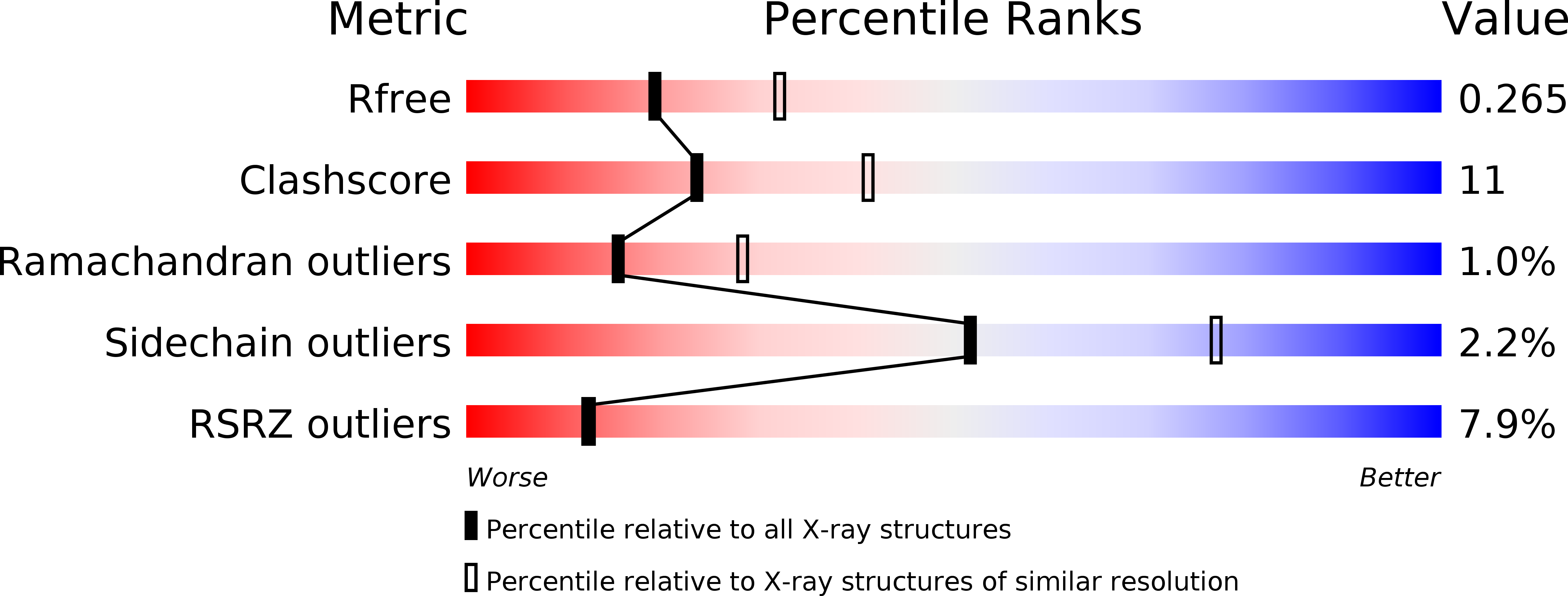
R-Value Free:
0.27
R-Value Work:
0.22
Space Group:
P 1 21 1