1OS7 image
Deposition Date 2003-03-18
Release Date 2003-09-23
Last Version Date 2024-02-14
Entry Detail
PDB ID:
1OS7
Keywords:
Title:
Crystal structure of TauD with iron, alpha-ketoglutarate and Taurine bound at pH 7.5
Biological Source:
Source Organism:
Escherichia coli (Taxon ID: 562)
Host Organism:
Method Details:
Experimental Method:
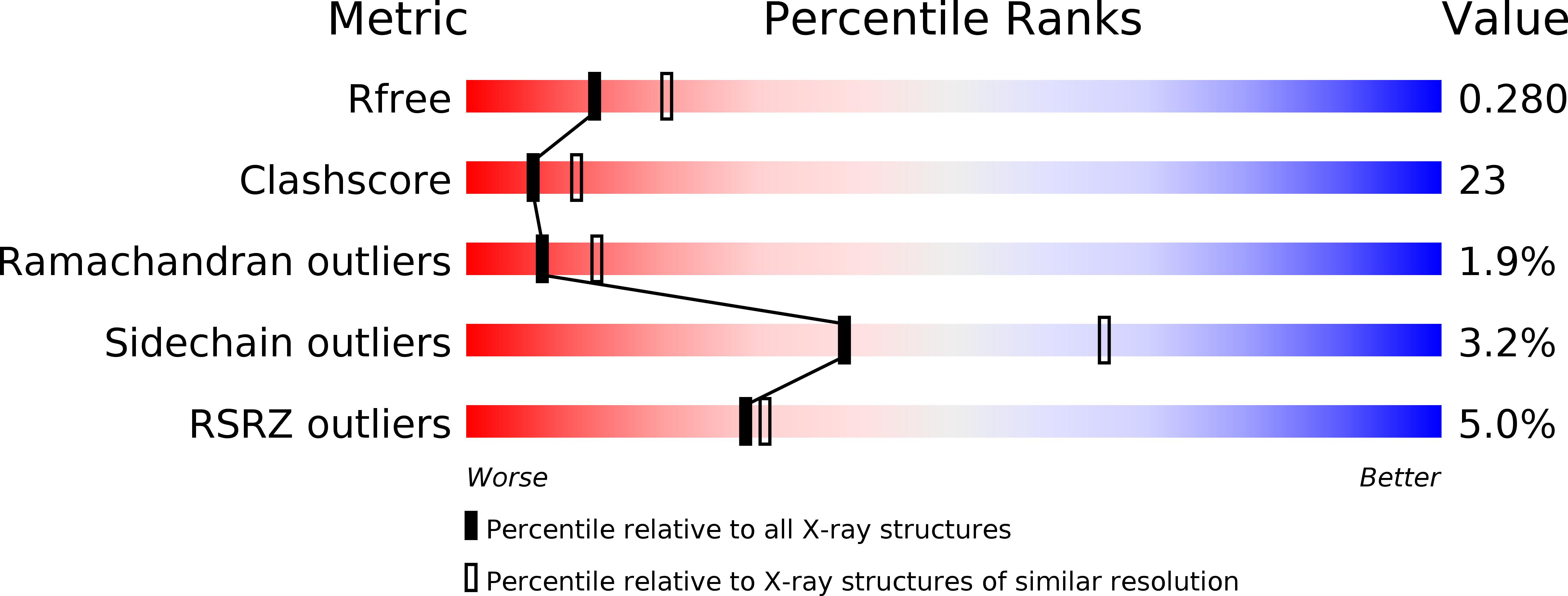
Resolution:
2.50 Å
R-Value Free:
0.27
R-Value Work:
0.22
R-Value Observed:
0.22
Space Group:
P 21 21 21
Macromolecular Entities
Structures with similar UniProt ID
Protein Blast
Polymer Type:polypeptide(L)
Molecule:Alpha-ketoglutarate-dependent taurine dioxygenase
Gene (Uniprot):tauD
Chain IDs:A, B, C, D
Chain Length:283
Number of Molecules:4
Biological Source:Escherichia coli
Primary Citation
Substrate-Induced Conformational Changes in Escherichia coli Taurine/alpha-Ketoglutarate Dioxygenase and Insight Into the Oligomeric Structure
Biochemistry 42 5547 5554 (2003)
PMID: 12741810 DOI: 10.1021/bi0341096

Abstact

The enzymes in the alpha-ketoglutarate (alphaKG) dependent dioxygenase superfamily represent the largest class of non-heme iron oxidases and have important medical, ecological, and biotechnological roles. One such enzyme, taurine/alpha-ketoglutarate dioxygenase (TauD), catalyzes the conversion of 2-aminoethanesulfonate (taurine) to sulfite and aminoacetaldehyde while decomposing alphaKG to succinate and CO(2). This alphaKG dependent dioxygenase is expressed in Escherichia coli under sulfur starvation conditions and allows the cell to utilize taurine, and other similar sulfonates in the environment, as an alternative sulfur source. In this work, we report the structures of the apo and holo forms of TauD to 1.9 A resolution (R(cryst) = 21.2%, R(free) = 24.9%) and 2.5 A resolution (R(cryst) = 22.5%, R(free) = 27.8%), respectively. The models reported herein provide significant new insight into the substrate orientations at the active site and the conformational changes that are induced upon taurine binding. Furthermore, analysis of our crystallographic data coupled with reanalysis of the crystallographic model (resolution = 3.0 A, R(cryst) = 28.1, R(free) = 32.0) presented by Elkins et al. (Biochemistry (2002) 41, 5185-5192) reveals an alternative oligomeric arrangement for the enzyme that is consistent with the conserved primary and secondary structure elements of other alphaKG dependent dioxygenases.

Legend

Protein

Chemical

Disease

Primary Citation of related structures