
Deposition Date
2003-03-18
Release Date
2003-09-23
Last Version Date
2023-11-15
Entry Detail
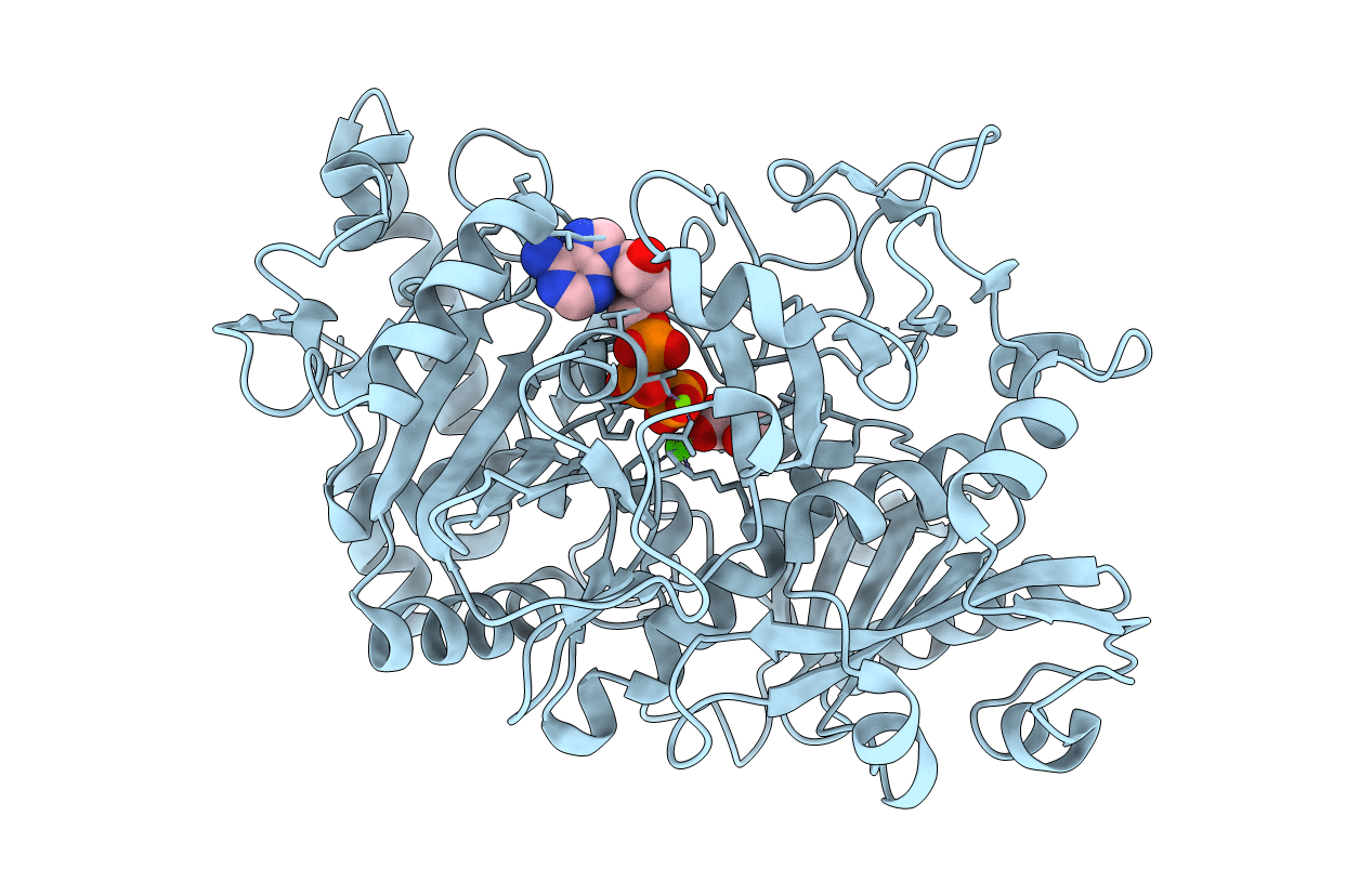
PDB ID:
1OS1
Keywords:
Title:
Structure of Phosphoenolpyruvate Carboxykinase complexed with ATP,Mg, Ca and pyruvate.
Biological Source:
Source Organism(s):
Escherichia coli (Taxon ID: 83333)
Method Details:
Experimental Method:
Resolution:
1.80 Å
R-Value Free:
0.25
R-Value Work:
0.2
R-Value Observed:
0.20
Space Group:
C 1 2 1
