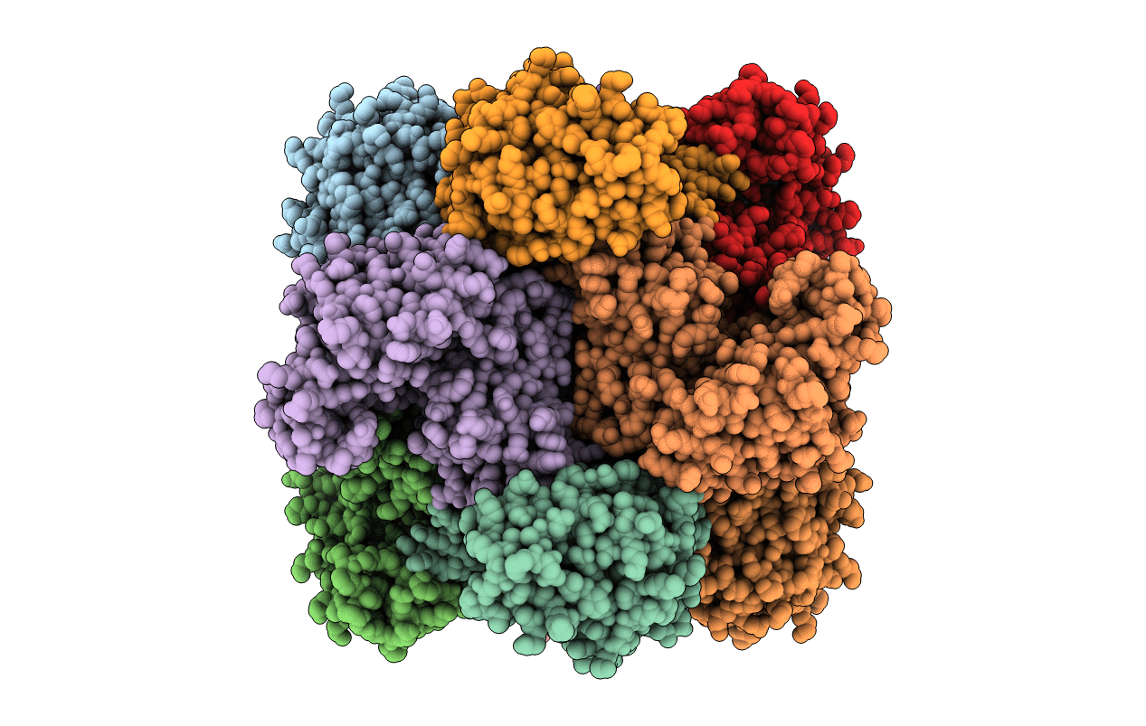
1ORT image
Deposition Date 1995-08-24
Release Date 1996-12-07
Last Version Date 2024-02-14
Entry Detail
PDB ID:
1ORT
Keywords:
Title:
ORNITHINE TRANSCARBAMOYLASE FROM PSEUDOMONAS AERUGINOSA
Biological Source:
Source Organism:
Method Details:
Experimental Method:
Resolution:
3.00 Å
R-Value Work:
0.21
R-Value Observed:
0.21
Space Group:
P 1
Macromolecular Entities
Structures with similar UniProt ID
Protein Blast
Polymer Type:polypeptide(L)
Molecule:ORNITHINE TRANSCARBAMOYLASE
Gene (Uniprot):arcB
Mutagens:E105G
Chain IDs:A, B, C, D, E, F, G, H, I, J, K, L
Chain Length:335
Number of Molecules:12
Biological Source:Pseudomonas aeruginosa
Ligand Molecules
Primary Citation
Crystal structure of Pseudomonas aeruginosa catabolic ornithine transcarbamoylase at 3.0-A resolution: a different oligomeric organization in the transcarbamoylase family.
Proc.Natl.Acad.Sci.USA 92 10762 10766 (1995)
PMID: 7479879 DOI: 10.1073/pnas.92.23.10762

Abstact

The crystal structure of the Glu-105-->Gly mutant of catabolic ornithine transcarbamoylase (OTCase; carbamoyl phosphate + L-ornithine = orthophosphate + L-citrulline, EC 2.1.3.3) from Pseudomonas aeruginosa has been determined at 3.0-A resolution. This mutant is blocked in the active R (relaxed) state. The structure was solved by the molecular replacement method, starting from a crude molecular model built from a trimer of the catalytic subunit of another transcarbamoylase, the extensively studied aspartate transcarbamoylase (ATCase) from Escherichia coli. This model was used to generate initial low-resolution phases at 8-A resolution, which were extended to 3-A by noncrystallographic symmetry averaging. Four phase extensions were required to obtain an electron density map of very high quality from which the final model was built. The structure, including 4020 residues, has been refined to 3-A, and the current crystallographic R value is 0.216. No solvent molecules have been added to the model. The catabolic OTCase is a dodecamer composed of four trimers organized in a tetrahedral manner. Each monomer is composed of two domains. The carbamoyl phosphate binding domain shows a strong structural homology with the equivalent ATCase part. In contrast, the other domain, mainly implicated in the binding of the second substrate (ornithine for OTCase and aspartate for ATCase) is poorly conserved. The quaternary structures of these two allosteric transcarbamoylases are quite divergent: the E. coli ATCase has pseudo-32 point-group symmetry, with six catalytic and six regulatory chains; the catabolic OTCase has 23 point-group symmetry and only catalytic chains. However, both enzymes display homotropic and heterotropic cooperativity.

Legend

Protein

Chemical

Disease

Primary Citation of related structures